- Autosomal dominant (AD)
- Autosomal recessive (AR)
- Sex-linked dominant
- Sex-linked recessive
- Multiple inheritance patterns
- Additional inheritance effects:
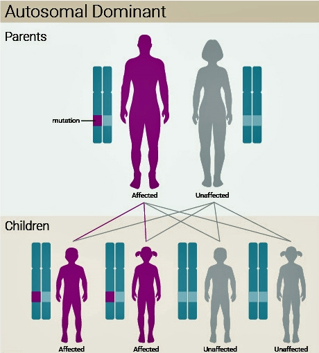
Autosomal dominant (Figure 1)
- A mutation in one copy of an autosomal gene causes the disease.
- A person with such a disease has one mutated and one normal copy of the disease gene.
- Risk of 1 in 2 to pass on to offspring.
- Variable expressivity (severity of the disorder) and incomplete penetrance(probability of a person with a mutation manifesting signs of the disease) suppress or minimise the expression of dominant inheritance.
- Autosomal dominant conditions usually produce structural abnormalities.

Figure 1. Autosomal Dominance Inheritance US National Library
Examples
- Syndactyly
- Polydactyly
- Marfan’s syndrome
- Cleidocranial dysostosis
- Hereditary multiple exostosis
- Achondroplasia
- Multiple epiphyseal dysplasia (MED)
- Metaphyseal chondrodysplasia (Schmid and Jansen types)
- Kniest dysplasia
- Malignant hyperthermia
- Ehlers–Danlos syndrome
- Osteogenesis imperfecta (types I and IV)
- Multiple hereditary exostosis
- Osteopetrosis (tarda, mild form)
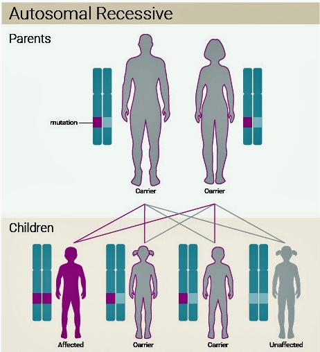
Autosomal recessive (Figure 2)
- Two copies of an abnormal gene must be present in order for the disease or trait to develop.
- A person with a recessive mutation in one copy and a normal second copy is a carrier or heterozygote.
- Both parents of a patient with an autosomal recessive disease are always carriers.
- Usually the parents do not know they are carriers until the birth of the first affected.
- The risk on future children with the disease (recurrence risk) is 1 in 4 for each child.
- Each child has a 25% chance of being affected, a 50% chance of being a carrier and a 25% chance of being normal.
- Autosomal recessive conditions are often metabolic or enzyme defects.

Figure 2. Autosomal Recessive Inheritance US National Library
Examples
- Diastrophic dysplasia
- Friedreich’s ataxia
- Gaucher disease
- Spinal muscular atrophy
- Sickle cell anaemia
- Osteogenesis imperfecta (II and III)
- Hypophosphatasia
- Osteopetrosis (infantile, malignant form)
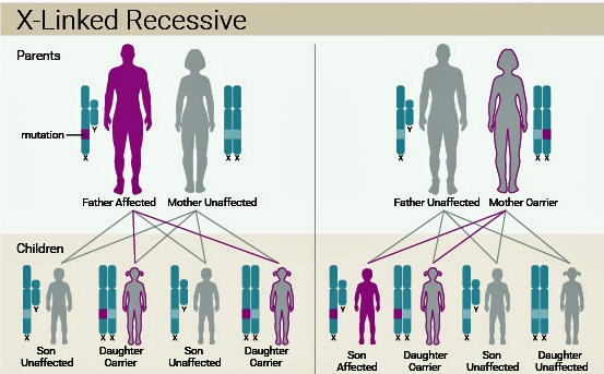
X-Linked recessive (Figure 3)
- Most of the X-linked recessive diseases are in men as they have only one X chromosome (XY).
- Women possessing one X-linked recessive mutation are considered carriers and will generally not manifest clinical symptoms of the disorder.
- All offspring of a carrier woman have a 50% chance of inheriting the mutation if the father does not carry the recessive allele.
- All female children of an affected father will be carriers (assuming the mother is not affected or a carrier), as daughters possess their father’s X chromosome.
- No male children of an affected father will be affected, as sons only inherit their father’s Y chromosome.
- In recessive X-linked inheritance, the woman is affected only in the rare situation in which both genes of the genetic pair are abnormal.

Figure 3. Autosomal Recessive Inheritance US National Library
Examples
- Duchenne muscular dystrophy
- Becker’s muscular dystrophy
- Mucopolysaccaridoses type II (Hunter's syndrome)
- Haemophilia A. Genetic defect in factor VII
- Spondylo-epiphyseal dysplasia (SED) tarda
Example of X-linked recessive condition
Duchenne muscular dystrophy
- Incidence approximately one in 4000 boys.
- This is one of the dystrophinopathies caused by a mutation in the dystrophin gene (Xp21).
- The dystrophin protein provides structural stability to the dystroglycan complex of the muscle cell membrane, which is lost as a result of the mutation. Women rarely show musculoskeletal signs of the disease, although there is an increased risk of dilated cardiomyopathy in female carriers.
- There is a high spontaneous new mutation rate.
Clinical features
- Age of onset is usually before 6 years. Progressive proximal myopathy of the lower limbs with noticeable calf pseudohypertrophy.
- Compensatory toe walking is an adaptation to knee extensor weakness.
- Frequent falls/fatigue.
- Speech delay and difficulty with motor skills with learning difficulties in approximately a third of affected boys.
- Lumbar lordosis/scoliosis.
- Usually wheelchair bound by 12 years and life expectancy is around 25 years.
- Gower’s sign positive: the child is unable to jump up quickly from a crossed leg position without bracing their arms against their legs to support the proximally weak muscles.
X-Linked dominant
- Rare.
- Each child of a mother affected with an X-linked dominant trait has a 50% chance of inheriting the mutation and thus being affected with the disorder.
- If only the father is affected, 100% of the daughters will be affected, because they inherit their father's X chromosome, and 0% of the sons will be affected, because they inherit their father's Y chromosome. This distinguishes its inheritance from autosomal dominant.
- Men are usuallymore severely affected than women because in each affected woman there is one normal allele producing a normal gene product and one mutant allele producing the non-functioning product, while in each affected man there is only the mutant allele with its non-functioning product and the Y chromosome, no normal gene product at all.
- Some X-linked dominant alleles are lethal in men because their sole X chromosome is the disease-carrying chromosome. In the next generation, sons of an affected father are unaffected because their X chromosome is from their mother. Daughters of an affected father are always affected as one of their chromosomes is inherited from their father.
Examples
- Hypophosphatemic rickets
- Leri–Weill dyschondrosteosis (bilateral Madelung's deformity)
Multiple inheritance patterns
Examples
- Charcot-Marie-Tooth (AD, AR, Xlink)
- Osteopetrosis (AD, AR)
- Osteogenesis imperfecta (AR, AD)
- Neurofibromatosis (AD, AR)
- SED (AD, Xlink)