TOPIC DETAILS

  Team Member Role(s) Profile
Fazal Ali Fazal Ali Section Editor, Segment Author
Mark Malek Racy Segment Author

Section editor: Fazal Ali

Segment author: Malek Racy and Fazal Ali

Document history: Version 1

Introduction 

Key points to elicit in the history:

  • Injury details e.g. knife or crush
  • Duration of chronic symptoms
  • Temporal pattern; worse during the day or night (nerve compression often worse at night)
  • Comorbidities

           - Diabetes
           - Thyroid disease
           - Other endocrine disease

  • Smoking/ alcohol

All three peripheral nerves in the upper limb have sensory and motor functions which can be tested to identify the location of pathology. Proximal and distal lesions are often referred to as ‘high’ and ‘low’ respectively. The examination technique is aimed at identifying this level in order to arrive at a diagnosis and guide management.

The order of examination is as follows:

  • Inspection
  • Sensation
  • Palpation
  • Power/ motor function

            - Proximally innervated muscles first moving distal

  • Provocative tests

             - Can be uncomfortable so do these at the end

Testing of the nerves is only possible if the anatomy is known:

Median nerve

Anatomy:

  • Medial and lateral cords of brachial plexus (C5-T1)
  • Runs between FDS and FDP in the forearm

           - Supplies superficial and intermediate muscles of flexor compartment (except FCU)

  • Gives off two branches in forearm

            - Anterior interosseous nerve

                     - Supplies the deep muscles of flexor compartment (except ulnar half of FDP)

             - Palmar cutaneous branch

                     - Branches 3-5cm proximal to carpal tunnel and passes superficial to it
                     - Supplies sensation to radial aspect of palm

  • Enters hand through carpal tunnel
  • Branches after carpal tunnel

           - Recurrent motor branch

                - Thenar muscles

           - Digital cutaneous branch

                - Sensation to palmar side of radial 3.5 digits
                - Sensation to dorsum of thumb, index and middle fingers

            - Branches to lateral (radial) two lumbricals

Compression syndromes:

Pronator syndrome

  • Compression of the median nerve at one of 4 locations:

           - Ligament of Struthers
           - Lacertus fibrosus
           - Pronator teres (PT)

                 - Between ulnar and humeral heads

          - Flexor digitorum superficialis (FDS)

  • Motor and sensory

AIN syndrome

  • Compression at multiple sites including:

           - PT
           - FDS
           - Lacertus fibrosus

  • Purely motor

Carpal tunnel syndrome

  • Motor and sensory

Inspection

  • Thenar wasting
  • Carpal tunnel release scar
  • Hand of benediction

 - Only seen on attempting to make a fist
 - Index and middle finger MCPJs remain in extension
 - Loss of lateral two lumbricals
 - Unable to flex DIPJ to index and middle
 - Loss of FDP and FDS to index and middle fingers
 - Unopposed pull of extensor tendons
 - Able to flex ring and small fingers
 - FDP to these and lumbricals supplied by ulnar nerve
 - Signifies a high median nerve lesion (proximal to FDP innervation)

 Hand of benediction MN 1.jpg

Figure 1 Hand of benediction.Only seen on making a fist.Unable to flex index and middle finger MCP Joints s and IP Joints due to high median nerve lesion.

Sensation

  • Palmar cutaneous branch

           - Base of the thenar eminence (palmar cutaneous branch arises 3-5cm proximal to wrist crease so this area is spared in carpal tunnel syndrome

  • Digital cutaneous branch

           - Index finger tip sensation

Interpretation:

  • Pathology distal to wrist e.g. carpal tunnel syndrome

           - Thenar sensation preserved

  • Pathology proximal to wrist e.g. forearm compression/ injury

           - Sensation affected over thenar eminence and fingertip

 Sensation median copy.png

Figure 2. Areas for testing median nerve sensation (shaded)

A:

  • Palmar cutaneous branch
  • Preserved in low lesion

B:

  • Digital cutaneous branch
  • Lost in both high and low lesions

Palpation

May (rarely) have tenderness over proximal forearm/ compression site

Motor

The three muscle groups to test are:

  • Proximal (to the carpal tunnel)

- Flexor carpi radialis (FCR)
- Flexor digitorum superficialis (FDS)

  • Distal (to the carpal tunnel)

- Abductor pollicis brevis (APB)
- Opponens pollicis (OP)

  • Anterior interosseous nerve (AIN)

- Flexor digitorum profundus (FDP)
- Flexor pollicis longus (FPL)

Sequence for median nerve motor testing (proximal to distal)

- Palpate the muscle/ tendon as you test each muscle group (not always shown to demonstrate anatomy)

 FCR.png

Figure 3 Flexor carpi radialis (FCR).Flex wrist against resistance and palpate tendon

 FDS.JPG

Figure 4 Flexor digitorum superficialis (FDS)

APB.JPG

Figure 5. Abductor pollicis brevis (APB).Support hand and ask the patient to abduct against resistance. Feel APB as the most radial thenar muscle

OP.JPG 

Figure 6 Opponens pollicis (OP).Touch thumb to small fingertip and resist you pulling them apart

FDP-OP.JPG 

Figure 7 Flexor digitorum profundus (FDP)/ flexor pollicis longus (FPL).Make the ‘OK’ sign

AIN palsy.JPG  

Figure 8 AIN palsy.If the AIN is deficient, the patient cannot flex at the IP joints of the thumb and the index finger

Provocative tests

Pronator syndrome:

Compression under lacertus fibrosus

  • Flex the patient’s elbow and pronate the forearm
  • The patient supinates the forearm against resistance
  • This tightens the biceps tendon/ lacertus fibrosus, reproducing the pain

Compression under PT

  • Fully supinate the forearm and pronate the arm against resistance
  • This tightens pronator teres, reproducing the pain

Compression under FDS

  • Flex the PIPJ of the index finger against resistance
  • This tightens FDS, reproducing the pain

Carpal tunnel syndrome:

Phalen’s test

  • Flex the patient’s wrists fully
  • Assess the amount of time taken to reproduce the symptoms

Reverse Phalen’s test

  • As above but with the wrists fully dorsiflexed/ extended

Tinel’s test

  • Percuss the median nerve just proximal to the wrist crease (where the nerve enters the carpal tunnel

Radial Nerve

Anatomy:

  • Posterior cord of brachial plexus (C5-T1)
  • Runs around spiral groove of humerus

            - Supplies medial and lateral heads of triceps

  • Passes through lateral intermuscular septum

             - Supplies brachioradialis, ECRL (and lateral brachialis)

  • Divides at level of radiocapitellar joint line

            1 Deep branch

                 - Supplies ECRB

           - Passes through radial tunnel

           - Runs around radial neck deep to supinator, becoming posterior interosseous nerve (PIN)- C7/8

                  -  Superficial branch
                      ECU, EDC, EDM
                 -  Deep branch
                     APL, EPL, EPB, EIP

         2 Superficial branch/ superficial radial nerve (SRN)

            - Runs deep to brachioradialis
            - Sensation to radial dorsum of hand

Potential sites of injury/ compression:

Arm

  • Spiral fractures of humerus (traumatic or iatrogenic)

Elbow (can mimic tennis elbow)

  • Radial tunnel syndrome

           - Compression at sites along radial tunnel
           - Pain only- no motor dysfunction

  • PIN syndrome

           - Compression at same sites as radial tunnel syndrome
           - Pain and weakness
 Wrist

  • Wartenberg’s syndrome

           - Compression of SRN at wrist (e.g. tight watch band)
           - Sensory

  • Iatrogenic/ traumatic from distal radius fracture

Inspection

  • Wrist drop/ use of splints
  • Scars (including upper arm for evidence of humeral injury/ surgery)
  • Wasting of brachioradialis

Sensation

Dorsum of 1st web space (superficial branch)

 Sensation radial.png

Figure 9. Area to test superficial radial nerve sensation (shaded)

Palpation

  • Differentiate between lateral epicondylitis (tennis elbow) and radial tunnel syndrome

           - Radial tunnel syndrome tenderness more distal

Motor

Examine muscles in order of innervation from proximal to distal

  • Radial nerve:

           - Brachioradialis (BR)
           - Extensor carpi radialis longus (ECRL)

  • PIN:  

           - Extensor carpi ulnaris (ECU)
           - Extensor pollicis longus (EPL)
           - Extensor indicis proprius (EIP)

Interpretation:

  • Complete weakness of wrist extension (ECRL and ECU)

            - High radial nerve lesion

  • Weakness of ulnar deviation in wrist extension (ECU) but good power of radial deviation in wrist extension (ECRL)

            - Pathology distal to bifurcation of radial nerve

                  Radial tunnel syndrome
                  Injury between innervation of these muscles

  • When testing finger extension, ensure the MCPJs also extend. IPJ extension alone is effected by the lumbricals (median/ ulnar nerves) so can give a false positive result

Sequence for radial nerve motor testing (proximal to distal)

Palpate the muscle/ tendon as you test each muscle group (not always shown to demonstrate anatomy)
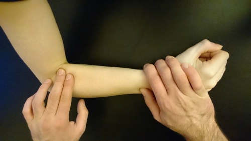
BR.JPG

Figure 10 Brachioradialis.With the elbow at 90° and the forearm in neutral pronation, flex the elbow against resistance and palpate the muscle
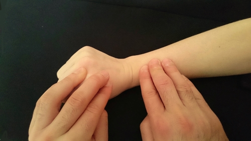
ECRL.jpg

Figure 11 ECRL.Extend and radially deviate wrist against resistance
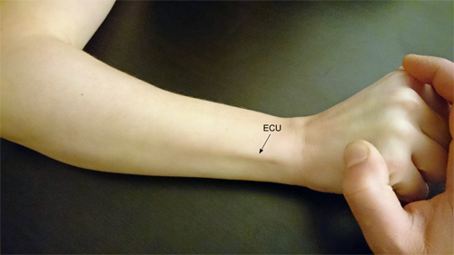
ECU.png

Figure 12 ECU.Extend and ulnarly deviate wrist against resistance
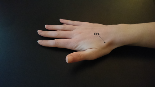
EPL.png

Figure 13 EPL.Thumb retropulsion

EIP.JPG

Figure 14 EIP .Point index finger with rest of fingers flexed to eliminate ED

Provocative tests 

PIN syndrome:

  • Extend patient’s elbow and resist active supination
  • This increases the pressure on the PIN (under the arcade of Frohse) causing pain in the forearm

Wartenberg’s syndrome

  • Tinel’s test along the superficial radial nerve

Ulnar nerve

Anatomy:

  • Medial cord of brachial plexus (C8/ T1)
  • Passes through medial intermuscular septum at arcade of Struthers
  • Runs behind medial epicondyle
  • Enters the forearm between humeral and ulnar heads of FCU

           - Supplies FCU and ulnar half of FDP (to ring and small fingers)

  • Divides

            1 Dorsal branch

               - 5cm proximal to wrist
               - Sensation from dorsum of ulnar border of hand and 1.5 fingers

             2 Palmar branch

                 - Enters hand through Guyon’s canal

                       - Superficial branch

                             - Sensation from palmar border of hand and 1.5 fingers

                       - Deep branch

                             - Interosseous muscles
                             - 3rd/ 4th lumbricals
                             - Adductor pollicis and medial head of flexor pollicis brevis
                             - Hypothenar muscles

Potential sites of injury/ compression:

Elbow

  • Points of compression along cubital tunnel:

            - Arcade of Struthers
            - Medial intermuscular septum
            - Medial epicondyle/ osteophytes
            - Osbourne’s ligament
            - Anconeus

Wrist

  • Guyon’s canal

Inspection

Wasting

  • Dorsal guttering
  • 1st dorsal interosseous
  • Hypothenar eminence

Scars

  • Guyon’s canal
  • Cubital tunnel

Wartenberg’s sign

  • Abducted and extended small finger

          - Weakness of 3rd palmar interosseous muscle
          - EDM (PIN) unopposed

Claw

  • Hyperextension of MCPJ and flexion of PIPJs of 4th and 5th fingers
  • Mechanism

             - 4th/ 5thlumbricals non-functioning (normally flex MCPJ and extend IPJs)
             - EDC causes hyperextension at MCPJ
             - FDP and FDS cause IPJ flexion

  • In a high ulnar nerve lesion, innervation to FDP is affected, causing reduced flexion of the ring and small fingers at DIPJ
  • The deformity looks less marked despite the injury being more severe
  • Known as the ‘ulnar paradox’

Wartenberg.JPG

Figure 15 Wartenberg’s sign

Ulnar claw.JPG

Figure 16 Ulnar claw.

  • Hyperextension of MCP Joints and flexion of IPJs 4th and 5th fingers
  • Image shown is a low ulnar nerve injury
  • In a high ulnar nerve injury, FDP is weak so there is no flexion of DIPJs 4th/ 5th fingers
  • The deformity looks less severe

Sensation

Dorsal cutaneous branch

  • Dorsum of small finger metacarpal

Superficial branch

  • Tip of small finger

Interpretation:

  • Proximal pathology e.g. cubital tunnel syndrome

            - Reduced sensation in both areas

  • Distal pathology e.g. Guyon’s canal

            - Sensation affected to tip of small finger
            - Sensation preserved dorsum of small finger metacarpal

Sensation ulnar.png

Figure 17 .Area to test ulnar nerve sensation (shaded)

A:

  • Dorsal cutaneous branch
  • Preserved in low lesion

B:

  • Superficial branch
  • Lost in a both high and low lesions

Palpation

Tender over cubital tunnel or Guyon’s canal

Take care not to confuse cubital tunnel tenderness with medial epicondylitis.

Motor

Examine muscles in order of innervation from proximal to distal

  • Flexor carpi ulnaris (FCU)
  • Flexor digitorum profundus (FDP) to small finger
  • Abductor digiti minimi (ADM)
  • First dorsal interosseous
  • (Froment’s test- see below)

Sequence for ulnar nerve motor testing (proximal to distal)

Palpate the muscle/ tendon as you test each muscle group (not always shown to demonstrate anatomy)

 FCU.png

Figure 18.  FCU.Flex and ulnarly deviate the wrist against resistance

FDP.JPG   

Figure 19. FDP to small finger.Flex DIPJ of small finger with FDS eliminated

 ADM.JPG

Figure 20.ADM.Abduct small finger against resistance whilst palpating muscle

1st dorsal interosseous.JPG 

Figure 21.1st dorsal interosseous.Abduct index finger against resistance whilst palpating muscle

Provocative tests

Tinel’s test over cubical tunnel/ Guyon’s canal 

Froment’s test

  • The patient should grasp a piece of paper in the 1st webspace/ between extended index finger and thumb
  • The examiner tries to pull the paper away
  • To resist requires adequate strength in the 1st dorsal interosseous muscle and adductor pollicis muscles (ulnar nerve)
  • To compensate and stop the paper from being pulled out of their hand, they flex the thumb using FPL (AIN)

 Froment -ve.JPG

Figure 22.Froment's test. Intact ulnar nerve

  • Full power in 1st dorsal interosseous/ adductor pollicis
  • Thumb extended and adducted to hold paper

PN 15.png

Figure 23.Froment's test.Ulnar nerve lesion

  • Weak 1st dorsal interosseous/ adductor pollicis
  • FPL used to grasp paper (AIN)

Quick testing

In certain situations, a quick neurovascular screening test may be appropriate (e.g. when assessing a child)

The three tests shown test gross median (AIN), radial and ulnar nerve motor function

Sensation can be assessed in the locations shown previously

FDP-OP.JPG

Figure 24.Median nerve (AIN).Make an ‘OK’ sign

 EIP.JPG

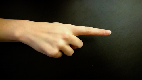
Figure 25 Radial nerve.Point index finger

Ulnar nerve.JPG 

Figure 26 Ulnar nerve.Cross fingers

Other peripheral nerves of the upper limb

 Axillary nerve

  • Supplies deltoid, teres minor and long head of triceps
  • Sensation:

            - Over the lateral deltoid (the ‘regimental badge’ area)

  • Motor:

            - Deltoid power
            - Passively abduct shoulder to 90°
            - Hold abducted against resistance/ downward pressure
            - Palpate deltoid for contraction (posterior/ central fibres)
            - Anterior fibres are tested as above but with the abducted shoulder in 90° of forward flexion

Musculocutaneous nerve

  • Supplies biceps brachii, brachialis and coracobrachialis
  • Sensation:

           - Lateral cutaneous nerve of the forearm (continuation of musculocutaneous nerve)

               - Sensation over lateral forearm

  • Motor:

            - Biceps brachii power
            - Adduct shoulder and flex elbow to 90°
             -Fully supinate forearm
            - Resist elbow flexion and palpate biceps

Previous
Next