TOPIC DETAILS

  Team Member Role(s) Profile
Fazal Ali Fazal Ali Section Editor
Gautum Gautam JK Tawari Segment Author

Section editor: Mr Fazal M Ali

Segment author: Mr Gautam JK Tawari

Document history: 10/9/2019

History

Age 

Age related likely conditions

  • Young patient – Instability
  • Middle age – Impingement, Adhesive capsulitis, Calcific Tendonitis, Rotator cuff tear.
  • Old Patient – Arthritis, Rotator cuff tear
  • Occupation
  • Handedness

Presenting Complaints

  • Pain
  • Weakness
  • Stiffness
  • Instability 

Functional and work related disability as a result of the presenting complaints should be sought  to provide an insight into the disability from the condition and helps treatment planning.  

Past Medical and Surgical history.

Medication, Allergies and Social history.

General Principles of Examination

  • Patients should be respected.
  • They must feel at ease.
  • Confidentiality & Dignity maintained.

Examination of shoulder joint 

  • Adequate exposure
  • Patient standing
  • Follow a systematic approach

  - Look, feel and move
  - Impingement Tests
  - Rotator Cuff assessment
  - Other Muscle assessment
  - Instability assessment
   -Neurological and vascular examination 

Look

General appearance:-

  • Syndromic features 
  • Congenital abnormalities
  • Orthopaedic stigmata
  • Horner’s Syndrome 

Neck and Shoulder Girdle:-

Inspection should be undertaken from the Front, side  and from behind. 

  • Attitude of the C-spine, shoulder/upper limb.
  • Bony Contours (SCJ, ACJ, Clavicle, Scapula and Shoulder joint)
  • Muscle Wasting

          - Deltoid (squared shoulder)
          - Supraspinous and Infraspinous scapular fossae  

  • Scars (Arthroscopy portals, Delto-pectoral area and in the Axilla)
  • Scapular position and winging
  • Popeye Sign (rupture long head of biceps)
  • Anterior axillary fold (rupture of Pectoralis Major)

Shoulder1.png

Figure 1. Inspection from front. Well hidden surgical scar 

Shoulder2.png

Figure 2. Inspection from side

shoulder3.png

Figure 3. Inspection from the back. Arthroscopy portal scar.

Screening for Neck Movements

  • Flexion (chin to chest)
  • Extension (looking up to the celling).
  • Lateral flexion (ear to shoulders)
  • Rotation (look side to side)

Restricted neck movements or reproduction of symptoms on neck movements indicate cervical spinal pathology.

Shoulder4.png

Figure 4 .Restricted neck extension

Shoulder5.png

Figure 5.Good neck flexion

Shoulder6.png

Figure 6. Restriction neck rotation 

Shoulder7.png

Figure 7.Restricted lateral flexion

Feel

  • Conform area of tenderness before beginning palpation

Systematic Palpation technique:

  • Sternoclavicular joint
  • Length of clavicle
  • Acromio-clavicular joint
  • Lateral border of acromion
  • Bicipital groove
  • Scapular spine (hollowness of Supraspinous and Infraspinous fossa should be felt at the same time)
  • Superior-medial scapular angle, medial scapular border & inferior scapular angle.  

Midline Cervical spine 

  • In the event of cervical spine pathology this should be performed to localise an approximate level of tenderness.

Shoulder8.png

Figure 8. Hollowness of supraspinatus fossa. 

Shoulder9.png

Shoulder10.png

Figures 9,10. Prominent posterior acromial spine and surgical scar.Wasting of posterior deltoid fibres.

Move

Movements of  the shoulder joint are a composite movement of Gleno-humeral joint (GHJ), Scapulo-thoracic joint (STJ), Acromio-clavicular (ACJ) and sternoclavicular joint (SCJ). The ratio of GHJ:STJ is 2:1.

Passive movements should follow ctive movements.

Active Movements 

Verbal instruction to elicit shoulder movements can be confusing; hence demonstration of movement required is preferred. 

All movements should be compared with the normal opposite side. 

Patients face must be observed at all times to help determine the comfortable range of active movements. 

  • Forward Flexion (FF)– 0°- 160°/170° (Normal)
  • Abduction (ABD)– 0°- 180° (Normal). Observation of abduction from behind the patient is important to determine scapular rhythm

Shoulder11.png

Figure 11.Demonstration of Active Forward Flexion

Shoulder12.png

Figure 12.Demonstration of Active Abduction.

Rotations – This can be measured in adduction & 90°of abduction. Hence it is important to specify the position of the arm in rotation.

  • External Rotation (ER) in Adduction (The arm must be on the side and the elbows flexed to 90°) – Variable hence must be compared.
  • Functional Internal Rotation (IR)– Reach of the extended thumb behind patients back. This can be determined as at greater trochanter, buttock, sacroiliac junction, L5-T12, Inferior scapular angle etc. 

Shoulder13.png

Figure 13. Demonstration of External rotation 

Shoulder14.png

Figure 14.Demonstration of Functional internal rotation

  •  External Rotation (ER) in 90°Abduction – The shoulder is abducted to 90°, elbow flexed to 90°and external rotation performed. It is generally more than internal rotation due to laxity of rotator interval & anterior superior capsule.
  • Internal Rotation (ER) in 90°Abduction – This is performed in the same position as above; the internal rotation is less than external rotation due to the taut postero-inferior capsule. 

Passive Movements

Performing passive movements is important to determine if the restriction of movement is due to – 

  • Pain – Movements can be increased some more but not without subjecting the patient to pain. Patients face should be observed at all times and the examination must be progressed with discreteness. 
  • Weakness – Movements can be increased significantly in case of weakness if not associated with pain.   

Shoulder15.png

Figure 15.Demonstration of Passive abduction

Shoulder16.png

Figure 16.Demonstration of Passive forward flexion

Impingement

Impingement pain is seen in an arc of 70°-120°. This is also known as the painful arc.

The tests for impingement bring the greater tuberosity underneath the acromion resulting in pain due to compression of the inflamed bursal tissue.

Impingement pain is not a diagnosis and an underlying cause should be sought. 

Neer’s Impingement Sign

  • The arm is internally rotated & passively elevated to >75°in the scapular plane. This elicits pain.

Neer’s Impingement test

  • In the event of a positive Neer’s sign, local anaesthetic is instilled in the subacromial space and the test reformed. The pain is relieved.

Hawkins-Kennedy test

  • The arm is brought in 90°of forward flexion, adduction and passively internally rotated. Presence of pain indicates impingement.

Shoulder17.png

Figure 17.Demonstration of Neer’s Sign. 

Shoulder18.png

Figure 18.Demonstration of Neer’s test. (post injection)

Shoulder19.png

Figure 19. Demonstration of Hawkins-Kennedy test

Yocum’s Test

  • The arm is adducted and the hand is rested on the opposite shoulder. Elbow is gradually elevated to horizontal plane. Presence of pain indicates impingement.

Shoulder20.png

Figure 20.Demonstration of Yocum’s test

Acromioclavicular Joint

Pathology of ACJ is suggested by localization of symptoms, joint tenderness & a high painful arc (>120°).

Scarf Test

  • Passive adduction of the arm across the midline so that the hand reaches towards the contra-lateral shoulder causes pain.

Shoulder21.png 

Figure 21.Demonstration of Scarf’s test

Shoulder22.png

Figure 22.Posteriorly prominent lateral end of clavicle on Scarf’s test.

Biceps Tendon

The long head of biceps inserts at the supraglenoid tubercle. Its role is not fully understood. Structural damage to this insertion is called a SLAP tear (Superior Labrum Anterior Posterior). The distal end of biceps inserts on the radial tuberosity at the elbow.

Proximal Biceps (Long Head)

  • Popeye Sign occurs on complete rupture of long head of biceps and is visible on inspection. 

Shoulder23.png

Figure 23.Demonstration of Popeye’s sign (Post surgical long head of biceps rupture)

Speed’s Test

  • The elbow is extended and the forearm supinated. Patient is asked to forward flex the humerus (like a ‘ten-pin bowling’ action). This movement is actively resisted. Pain is felt in the bicipital groove.

Shoulder24.png

Figure 24.Demonstration of Speed’s test

Yergasson’s Test

  • Patients hand is taken in ‘handshake’ position with elbow flexed at 90°and asked to pronate & supinate the forearm. This movement is resisted. Pain is present only on resisted supination.

Shoulder25.png

Figure 25. Demonstration of Yergasson’s test.

O’Brien Test

Shoulder forward flexed to 90°and adducted 15°. Downward force exerted with shoulder internally rotated (Thumb points down) and then externally rotated (Thumb points up). Pain worse on internal rotation is positive for SLAP lesion

Shoulder26.png 

Shoulder27.png

Figure 26 and 27. Demonstration of O’Brien’s Test

Distal Biceps

Examination of distal end of biceps is routinely performed as a part of elbow examination. But this should be examined in patients with biceps pathology.

Shoulder28.png 

Figure 28.Rupture distal end of Biceps

Palpation

  • The tendon can be easily palpated with elbow flexed at 90°and forearm fully supinated.

Hook test

  • Resisted flexion of elbow with forearm supination allows the distal biceps tendon to be taut. A finger can be easily wrapped around the tendon in shape of a hook and tension in the tendon tested.

Inability to hook the tendon indicates rupture.  

Shoulder29.png

Figure 29.Demonstration of Hook test for distal end of biceps (Normal)

Shoulder30.png

Figure 30.Demonstration of inability to perform a hook test in distal biceps rupture.

Rotator Cuff Assessment

The rotator cuff muscles should be tested in sequence followed by deltoid & other muscles of shoulder girdle.

Supraspinatus

  • This is an abductor of the shoulder. Deltoid also assists in abduction of the shoulder and is more powerful than supraspinatus.

Jobe’s ‘Empty Can’ Test

The shoulder is abducted to 90°in the scapular plane (30°forward flexion). The arm is internally rotated and forearm pronated so that the thumb points the ground. Push down and ask patient to resist. Feel the muscle.  
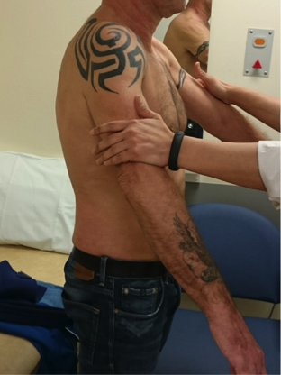
Shoulder31.png

Figure 31.Demonstration of Jobe’s test (Performed from front) 
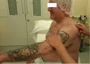
Shoulder32.png 

Figure 32.Demonstration of Jobe’s test (Performed from side) The muscle is felt.

Infraspinatus and Teres Minor

Infraspinatus is the most powerful external rotator of the shoulder when the arm is by the side. 

Testing Infraspinatus 

  • Elbow on the side and flexed at 90°. Ask the patient to externally rotate and resist this movement. Feel the muscle.

External Rotation Lag Sign

  • Elbow on the side and flexed at 90°. The shoulder is placed passively in full external rotation and patient asked to maintain this position. The hand holding the arm is released. The test is positive if the forearm drops back towards neutral position. (Infraspinatus tear)
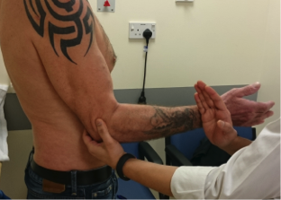
Shoulder33.png

Figure 33.Testing External rotation in adduction (Infraspinatus, Performed from front) 
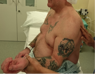
Shoulder34.png

Figure 34.Testing External rotation in adduction.(Infraspinatus, Performed from side) The muscle is felt.

Teres Minor

Teres minor assists infraspinatus in external rotation. Teres minor can be isolated by abduction of shoulder to 90°and testing external rotation.

Testing for Teres Minor

  • Hornblowers Sign-Shoulder abducted to 90°, elbow flexed to 90°and shoulder externally rotated. Patient can maintain this position even against some resistance. Inability to maintain this position results in results in hand falling in front of the face with further elbow flexion. 
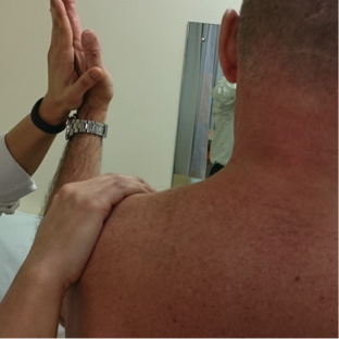
Shoulder35.png

Figure 35.Testing External rotation in abduction (Teres Minor isolated)

Subscapularis

It is a powerful internal rotator of the shoulder and forms the force couple with infraspinatus. 

Gerber’s Lift off Test

  • The arm is placed behind the back and the hand is positioned away from the body. Inability to maintain this position indicates muscle weakness.
  • Limitation - Patients with restricted internal rotation may struggle to get their hand behind their back. 
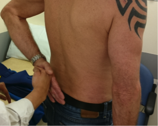
Shoulder36.png

Figure 36.Demonstration of Gerber’s Lift Off Test

Belly Press Test

  • The elbow is kept forward and the patient is asked to press their hand into their abdomen. 
  • This is an alternate test for subscapularis if shoulder movements are restricted. 
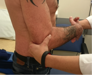
Shoulder37.png

Figure 37. Demonstration of Belly Press test.   

Other Muscles

The muscles around the shoulder girdle should be tested for their function and strength.

Deltoid, Pectoralis Major & Latissimus Dorsi are tested from the front of the patient. Trapezius, Rhomboids and Serratus anterior are test from behind the patient. 

From the front

Deltoid

  • Shoulder is passively abducted to 90°and extended. Patient is asked to maintain abduction against resistance. This tests posterior deltoid fibres.
  • Placing the abducted arm in neutral and flexed position respectively allows testing for central & anterior fibres.

Shoulder38.png

Figure 38.Testing Deltoid, comparing power and eliciting weakness.

Shoulder39.png

Figure 39.Testing Deltoid. The muscle is felt.

Pectoralis Major

  • Hand on waist and squeeze inwards. Palpate the muscle.

 Shoulder40.png

Figure 40.Testing pectoralis major. The muscle is felt.

Latissimus Dorsi

  • Downward and backward pressure of arm against resistance, as though climbing a ladder. Palpate the muscle. 

Shoulder41.png

Figure 41.Testing Latissimus Dorsi. The muscle is felt.

From Behind

Trapezius

  • Patients shrugs shoulders against resistance

Shoulder42.png

Figure 42.Testing trapezius. The muscle is felt.

Serratus Anterior

  • Patient is asked to push against a wall, fingers and palm pointing downwards. Winging of the scapula is noted.

Shoulder43.png

Figure 43.Testing serratus anterior. Winging of the scapula should be noticed if present.

Rhomboids

  • Hands on the hips and pushing the elbow back against resistance. Feel the muscle.

 Shoulder44.png

Figure 44.Testing Rhomboids. The muscle is felt.

Instability

Examination should systematically evaluate instability in each direction. Traumatic & Atraumatic causes overlap.

The tests for instability can be performed with patient sitting or lying down. 

Sulcus Sign

  • Passive downward traction on humerus in neutral position.
  • Determines presence of inferior laxity.
  • Positive sulcus sign seen in external rotation indicates incompetence of rotator interval structures.

Shoulder45.png

Figure 45.Demonstration of Sulcus Sign

Drawer Test (Load and Shift Test)

  • Anterior and Posterior Drawer test – It is performed by passively displacing the humeral head anteriorly or posteriorly. The scapula should be fixed with one hand and the other hand displaces the humeral head.

Shoulder46.png

Shoulder47.png

Figure 46 and 47.Demonstration of Anterior & Posterior Drawer Test (Load and Shift Test)

Apprehension Test

  • Anterior Apprehension Test- Abduction of shoulder to 90° with external rotation to 90° causes apprehension in the patient. Posteriorly directed pressure on the humeral head relieves the apprehension (Relocation Test)
  • Posterior Apprehension Test- The arm is held in forward flexion of 90°, adductionand internal rotation. The elbow is flexed to 90° and an axial load is applied along the humerus. The patient feels apprehension. Direct pressure on the humeral head relieves apprehension.

Shoulder48.png

Figure 48.Demonstration of Anterior apprehension Test (Patient Lying)

Shoulder49.png

Figure 49.Demonstration of Anterior apprehension Test (Patient sitting)

Shoulder50.png

Figure 50. Demonstration of Posterior apprehension Test (Patient Lying)

Shoulder51.png

Figure 51.Demonstration of Posterior apprehension Test (Patient sitting)

Generalised Ligamentous Laxity 

  • Beighton Score– This should be performed particularly in patients with bilateral symptoms or multidirectional instability. A total maximum score 9 can be obtained with a score greater than or equal to 4 indicating hypermobility.  

Thoracic outlet syndrome

Patients with thoracic outlet syndrome may present with shoulder symptoms. 

Presentation may include a combination of neurological, or vascular symptoms and fatigability of arm.

Physical examination of shoulder is more often than not normal.

Neurological Component Test

  • Roo’s Test– Brace the shoulders back fully, shoulder abducted to 90° and flex the elbows to 90°. Flex and extend fingers rapidly. Reproduction of symptoms confirms diagnosis.

 Vascular Component Test

  • Adson’s Test– Extend the head and rotate it to the affected side. Abduct the ipsilateral arm to 30° and feel the radial pulse. Patient is asked to take a deep breath and hold it. If the radial pulse disappears then the test is positive.

Shoulder52.png

Figure 52.Demonstration of Roo’s test. 

Shoulder53.png

Figure 53.Demonstration of Adson’s test. Loss of radial pulse when patient holds a deep breath.

CASE BASED DISCUSSIONS

Proximal Biceps rupture

Although proximal biceps rupture “Popeye sign”is a spot diagnosis candidates still need to know how to subsequently proceed with a diagnosis specific examination and have answers prepared for the likely questions that will be asked.

Popeye 1.jpg

Short case spot diagnosis

On inspection there is an obvious Popeye sign in the anterior flexor compartment of the arm suggestive of a LHB rupture. I would like to confirm my finding.

I would like to test for power of flexion and extension of the arm.

During active flexion the biceps retracts producing the classic deformity of the biceps contour

Perform :Speeds, Yergason’s and Ludington tests.

Jobe test positive. Confirms supraspinatous weakness. Rest of the rotator cuff examination normal.

Examine for lag signs.None present

Plan. Order MRI scan to look for a cuff tear and degree of fatty atrophy. Buzz words are “Goutallier grade

If a significant functional disability, consider supraspinatus repair and LHB tenodesis.

Discussion 

Proximal and distal attachments of the biceps tendon

Techniques for biceps repair both proximally and distally

Nerve supply and actions of the rotator cuff

Previous
Next