TOPIC DETAILS

  Team Member Role(s) Profile
Fazal Ali Fazal Ali Section Editor
James James Tomlinson Segment Author

Section editor: Mr Fazal Ali

Segment author: Mr James Tomlinson

Document history: 14/09/2019

General Questions

  • Age, Occupation, Hobbies
  • Treatment Goals: Relief of pain? Reassurance? Return to function? (this is important to establish as may have unrealistic aims which are not achievable)
  • Duration of symptoms
  • Previous treatment? (e.g. physio, injection, pain clinic, analgesics)
  • Ongoing litigation (needs careful phrasing)
  • Outcome scores (Now part of British Spine Registry - usually ODI/EQ5-D)
  • Past medical history
  • Drug history
  • Smoking (especially if considering fusion surgery) 

Age

  • <10 years 

Congenital/Infantile scoliosis

  • 10-20 years

Idiopathic scoliosis, Scheurmann’s disease, Lytic spond

  • Young adults

Ankylosing spondylitis (IBD?, Uveitis?), Disc disease

  • Middle age

Degenerative lumbar/cervical slip, Spinal stenosis, Discitis, Tumour

  • Elderly

Osteoporosis, Degenerative lumbar/cervical disease, Spinal stenosis, Tumour 

Pain

  • Leg vs Back (must distinguish between two)
  • Distribution (L3 - ant thigh, L4 anteromedial lower leg, L5 lateral calf, S1 posterior calf/sole of foot)
  • Neck vs Arm if cervical
  • Distribution (C5 lat upper arm, C6 lat forearm, C7 middle finger, C8 medial forearm, T1 medial upper arm)
  • Duration?
  • Onset? (rest, exercise)
  • Walking distance? (important in stenosis)
  • Relieving factors - Rest? Sitting down/Sitting forward? (Stenotic pain usually not relieved without sitting)
  • Walking up/down hill (Stenotic pain often easier uphill - c.f. vascular claudicant) 

Gait

  • Poor balance
  • Clumsiness
  • Weakness - unilateral vs bilateral
  • Walking aids? 

Bladder/Bowel

  • Essential part of spine history
  • Awareness of bladder filling?
  • Normal stream/control?
  • Incontinence? (Stress vs Urge)
  • Post void dribbling? 

Paediatric Deformity

  • Age
  • Age of onset
  • Birth history
  • Developmental milestones
  • Family history
  • Hyperlaxity
  • Main concern (e.g. rib hump, waist asymmetry, non-cosmetic concerns)
  • Menarchae
  • Other medical history (CP, DMD, Neurofibromatosis, Metabolic disorders, Chromosomal abnormality)
  • Pain
  • Progression of deformity
  • Rate of progression
  • Scoliosis outcome scores (SRS-22) 

Yellow Flags

  • Belief that symptoms treatable
  • Catastastrophisation
  • Compensation
  • Depression (does not exclude treatment)
  • Work issues? 

Red Flags

  • Age <16 years, >50 with back pain
  • Bilateral leg pain
  • Bladder dysfunction
  • Malignancy 
  • Severe infection
  • Steroids/Immunosupression
  • Thoracic back pain
  • Unplanned weight loss 


Inspection

General

  • Height, weight
  • Walking aids
  • Undress patient to see whole spine
  • Stigmata of spine disease (café au lait, axillary freckling, hair tuft/dimple, foot deformity)
  • Hyperlaxity (Beighton score)

Inspection 

Standing

Spinal Balance

  • Coronal profile 
  • Head centered over pelvis (plumbline)
  • Scars
  • Shoulder height
  • Rib hump (Forward bend test)
  • Waist asymmetry
  • Pelvic tilt
  • Leg length

 Sagittal profile

  • Cervical/Lumbar lordosis
  • Thoracic kyphosis
  • Head over pelvis

Clinical spine 1.png

Figure 1.Clinical picture showing C7 plumb line with normal alignment.  If Plumb line does not hang over pelvic midline suggests coronal plane deformity.

Standing

Limbs

  • Muscle wasting
  • Fasiculation
  • Cutaneous pigmentation (vascular disease)

Inspection

Standing 

Screening tests

This allows a rapid screening of lower limb myotomes before formal assessment

  • Trendelenburg (L5 myotome)
  • Single leg squat (L3)
  • Heel stand (L4)

Tip toe stand (S1)

Clinical spine 2.png

Figure 2. Close up shot to show single leg stance.  Test L5 by feeling pelvis and asking patient to repeat this.  If weak pelvis falls to non WB side 

Clinical spine 3.png

Figure 3.Close up shot to show single leg squat. Tests L3 myotome.

Clinical spine 4.png

Figure 4.Close up shot to show heel stance.  Tests L4 myotome.

This position is difficult to hold – alternative is to ask the patient to walk on their heels.

Palpation

Standing

  • Spinous processes
  • Paraspinal muscles
  • Localised raised temperature
  • Sacroiliac joint tenderness

Movement

Standing

Cervical spine

  • Flexion
  • Extension
  • Rotation
  • Lateral Flexion 

Thoracic spine

  • Rotation (limited value, and do with patient seated to fix pelvis) 

Lumbar spine

  • Flexion (Level reached by finger tips)
  • Schober’s test

(Mark L5 spinous process, then draw line 5cm below and 10cm above this) The line should increase by >5cm on forward flexion of L-spine.  If abnormal may indicate Ankylosing spondylitis.  

  • Extension

Lateral flexion 

Clinical spine 5.png

Figure 5.Lumbar flexion.In this case it would be documented as ‘lumbar flexion finger tips to floor’

Clinical spine 6.png

Figure 6.Shobers test with lines drawn – 5cm below and 10cm above. (not to scale)

Clinical spine 7.png

Figure 7.Shobers test being performed.  Must remeasure distance between top and bottom lines and should have increased >5cm. (i.e. by 20cm or more in total)  (The measurements here are from pre test marking.)

Clinical spine 8.png

Figure 8.Lumbar extension.  Limited value but should be assessed.

Clinical spine 9.png

Figure 9.Lateral flexion. (left side)  Document as ‘finger tips to mid thigh’ and repeat on right side.

Inspection

Walking

Gait

  • Normal
  • Antalgic 
  • Wide based (Myelopathy?)
  • Foot drop (L4?)
  • Trendelenburg (L5?)

Neurological assessment

Sitting

Upper Limbs

Tone, sensation, power, reflexes and peripheral pulses should be checked.

Tone

  • ‘Shake hands’ and rotate forearm and flex/extend elbow 

Sensation

Assess light touch and sharp sensation routinely.  Temperature (spinothalamic tract), vibration sense and two-point discrimination (dorsal columns) are useful for detailed examination. 

Dermatomes: 

  • C5 (lateral upper arm)
  • C6 (thumb)
  • C7 (middle finger)
  • C8 (little finger)
  • T1 (medial upper arm) 

Power

Graded 0-5 using MRC scale. 

Myotomes:

  • C5 (Shoulder abduction)
  • C6 (Elbow flexion)
  • C7 (Wrist extension)
  • C8 (Finger flexion)
  • T1 (Finger abduction) 

Note: There is sometimes disagreement on upper limb myotomes between clinicians.  It may be better to say ‘Shoulder abduction is 5/5, as this is more easily repeatable without ambiguity.

MRC scale

  • 0 No movement
  • 1 Flicker of contraction
  • 2 Movement with gravity eliminated
  • 3 Movement against gravity
  • 4 Movement against resistance but reduced
  • 5 Normal power 

Reflexes

  • Present, Reduced, Brisk, Absent
  • Biceps (C5)
  • Bracioradialis (C6)
  • Triceps (C7) 

Clinical spine 10.png

Figure 10.Upper arm dermatomes. Test C5 over lateral upper arm, T1 inner upper arm. Note: Different texts use differing dermatomal maps – may be some disagreement.

Clinical spine 11.png

Figure 11.Hand dermatomes.Test C6 palmar aspect thumb, C7 palmar middle finger, C8 palmar little finger

Clinical spine 12.png

Figure 12.C5 myotome.Resisted abduction.  Press down on arm while palpating deltoid to feel muscle contraction.

Clinical spine 13.png

Figure 13.C6 myotome.Resisted elbow flexion.Palpate biceps to feel muscle contraction.

Clinical spine 14.png

Figuure 14.C7 myotome.Resisted wrist extension. Palpate common extensor origin to feel contraction.

Clinical spine 15.png

Figure 15.C8 myotome.Resisted finger flexion.

Clinical spine 16.png

Figure 16.T1 mytome.Resisted finger abduction

If any suggestion of brisk reflexes perform Hoffman’s test. (flexion of middle finger DIPJ - thumb IPJ flexion if positive).  If positive suggests corticospinal tract abnormality and possible myelopathy 

Clinical spine 17.png

Figure 17 Hofman reflex. Flex IPJ index finger and watch for thumb IPJ flexion.

Pulses

  • Brachial
  • Radial
  • Ulna 


Special tests

Special or provocative tests should be done at the end of the examination as they may provoke pain.  

(If performing lower limb examination then perform special tests after this)

 Spurlings test

  • Lateral flexion to symptomatic side with neck extension
  • Positive if pain in ipsilateral arm within 30 seconds
  • If initially negative apply axial load on cranium as further provocation 

(Specific for foraminal compression but not highly sensitive)


 L’hermitte Sign 

  • Cervical spine flexion and/or extension precipitates shock like pain in arms or legs 

(Specific for myelopathy but not highly sensitive) 

Hoffman test

Hold middle phalanx of index or middle finger

Flick distal phalanx of finger
Positive test is flexion of thumb IPJ
 
(Sensitive for cervical myelopathy but not specific)

Neurological assessment lower limbs

Lying 

Lower Limbs 

Tone, sensation, power, reflexes and peripheral pulses should be checked. 

Tone 

Roll leg on examination couch – assesses tone and hip pain. 

If tone normal foot moves out of sequence with leg/knee.  If tone normal foot moves with leg. 

Sensation 

Assess light touch and sharp sensation routinely.  Temperature, vibration sense and two-point discrimination are useful for detailed examination. 

Dermatomes

  • L1 (groin)
  • L2 (anterior thigh)
  • L3 (anterior knee)
  • L4 (anteromedial lower leg)
  • L5 (lateral calf)
  • S1 (sole of foot) 

Clinical spine 18.png

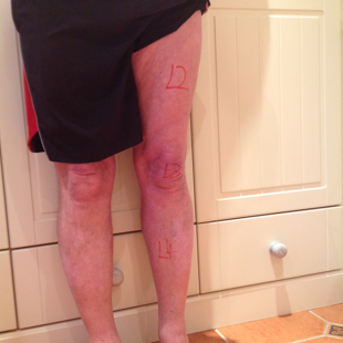
Figure 18.Leg dermatomes.L2 tested over anterior thigh, L3 over knee and L4 over medial lower leg.Note: Different texts use differing dermatomal maps – may be some disagreement.
Clinical spine 19.png

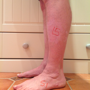
Figure 19.Foot dermatomes.L5 tested over lateral calf and dorsum of foot.S1 tested over lateral border of foot/sole of foot. 

Power

Graded 0-5 using MRC scale. 

Myotomes

  • L2 (hip flexion)
  • L3 (knee extension)
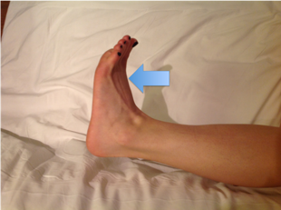
  • L4 (foot dorsiflexion)
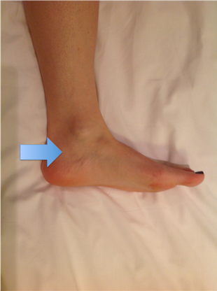
  • L5 (great toe dorsiflexion)
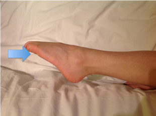
  • S1 (foot plantarflexion) 

Note: Although there is far less variation in description of lower limb myotomes than upper limb it may still be helpful to describe as ‘hip flexion was 5/5’ or ‘knee extension was 4/5’. 

Reflexes

  • Present, Reduced, Brisk, Absent
  • Quadriceps (L3/4) (to relax the quads for optimal testing ask the patient to sit on couch with legs hanging free)
  • Ankle (S1)
  • Plantars – rub lateral sole of foot travelling distally and across metatarsal heads
  • Downgoing/Equivocal plantar reflex is a normal response

Upgoing plantar reflex suggests upper motor neurone pathology – either spinal cord or brain 

Note: If up-going plantars and not previously identified then consider brain/spinal cord MRI. 

If reflexes brisk then examine for clonus - >3 beats is abnormal.   

(If reflexes normal do not assess for clonus) 
Clinical spine 20.png

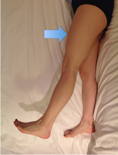
Figure 20.L2 myotome.To test against resistance apply downward pressure over mid femur (reduces mechanical advantage of tester).If power unable to resist gravity ask patient to lie on side and repeat 
Clinical spine 21.png

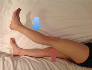
Figure 21.L3 myotome.Support leg just above knee (red arrow) and then test knee extension with downward pressure on lower leg (blue arrow)
Clinical spine 22.png

Figure 22.L4 myotome.Ask patient to dorsiflex ankle and apply resistance over forefoot as shown by blue arrow.
Clinical spine 25.png

Figure 23. L5 myotome.Ask patient to extend great toe and apply resistance over great toe as shown by blue arrow.
Clinical spine 24.png

Figure 24. S1 myotome.Ask patient to plantarflex foot and apply resistance under MT heads as shown by blue arrow.

Pulses

  • Posterior tibial
  • Dorsalis pedis

If abnormal consider vascular opinion – especially if claudicant symptoms.

Clinical spine 25.png

Figure 25.Blue arrow shows approximate location of posterior tibial pulse.It is found posterior and inferior to the medial malleolus

Clinical spine 26.png

Figure 26.Blue arrow shows approximate location of dorsalis pedis pulse.It is found lateral to the EHL tendon

Special tests

Special or provocative tests should be done at the end of the examination as they may provoke pain.  

 Lasegue’s test/sign 

  • Patient supine
  • Elevate leg with knee extended
  • If pain in posterior thigh/calf then lower leg and dorsiflex ankle
  • Pain on dorsiflexion = positive test

Bowstring test

  • Patient supine
  • Elevate leg with knee extended
  • If pain in posterior thigh/calf then lower leg and apply pressure in popliteal fossa
  • Pain on pressure = positive test

Crossover test

  • Patient supine

Elevate leg on unaffected side

  • Pain on affected side = positive test

Suggests more severe root irritation but does not correlate with scan findings reliably

Further examination

PR 

  • Sensation
  • Tone 

This is important to complete any spinal examination. Mention it but usually not performed in exam setting. 

Other examination

Waddell described five signs which he suggested may help distinguish between pain or organic and non-organic pathology. 

The reliability of these signs is sometimes disputed, and it is unlikely you will be asked to demonstrate them.  You should have an awareness of them though. 

Waddell’s signs

  • Non anatomic tenderness
  • Pain on axial loading
  • Distraction test (i.e. positive test negative when patient distracted)
  • Non dermatomal pain or sensory change
  • Overreaction 

Paediatric Spine (Deformity)

Follow same system as above with few key additions as per below: 

Inspection 

Stigmata of disease 

  • Café-au-lait spots, axillary freckling, neurofibromas
  • Blue sclera, dental abnormality (Osteogenesis imperfecta)
  • Pectus carinatum/excavatum, high arched palate, hyperlaxity (Marfans)
  • Hair tuft, sacral dimple (Spina bifida) 

Leg length discrepancy

  • Many possible causes
  • See pelvic tilt and thus may see secondary scoliosis 

Coronal balance

  • C7 plumb line
  • Palpate C7 spinous process 
  • Hang plumb line from this level

Should be centered over pelvis 

Adams forward bend test

  • Patient standing
  • Bend forward from waist
  • Look for rib hump – asymmetry of thoracic cage

Sign of rotational deformity – differentiates structural scoliosis from non structural 

Clinical spine 27.png

Figure 27.Adams forward bend test.This is a normal spine so no asymmetry.If positive see asymmetry of thoracic cage.

Palpation 

Same as adult spine examination above 

Movement

Same as adult spine examination above 

Neurological assessment

Same as adult spine examination except abdominal reflexes. 

Reflexes

Abdominal 

  • Gently scratch four quadrants of abdomen
  • See contraction of abdominal musculature toward each quadrant
  • Asymmetry may suggest dysraphism 

Note: Abdominal reflex may be absent due to obesity or lax musculature. 

May be abnormal in MS, MND, Brown-Sequard as well as dysraphism.  

Previous
Next