TOPIC DETAILS

No contributor information available.

Metatarsus Adductus

  • It is the commonest foot deformity in pre-toddlers
  • The forefoot is adducted relative to the hindfoot
  • The deformity presents either at birth or shortly thereafter
  • Sole of the foot may have a kidney bean shape

Etiology

  • It is thought to be a packaging disorder
  • MA is associated with torticollis, DDH etc
  • MA is only seen in term babies but not in pre-term
  • Idiopathic MA has an incidence of 3%

Pathoanatomy

  •  It is thought that primary abnormality is at the medial cueniform.
  • Medial cuneiform articular surface is medially deviated
  • Others have claimed that the apex of angulation is at the Lisfranc joint and there is subluxation at the tarso-metatarsal (MTT)
  • Metatarsal metaphyses are adducted
  • Actively correctable: stroking the lateral border of the foot leads to active correction
  • Passively correctable: foot can be passively corrected
  • Rigid: these have deep medial crease and are not correctable
  • Parents may notice in toeing if child presents after walking age
  • Forefoot is adducted relative to the hindfoot
  • Viewed from the plantar surface the sole of the foot appears like a bean
  • Lateral border of the foot is convex
  • Medial border of the foot is concave
  • Base of the 5th metatarsal may be more prominent than normal
  • Space between the 1st and 2nd toe is also increased
  • Ankle and sub-talar joints’ movement is normal
  • It is important to undertake a thorough clinical assessment to rule out plagiocephaly, torticollis and DDH
  • It is also important to assess lower limb rotational profile to rule out other causes of internal rotation of the foot
  • However MA may co-exist with a degree of Internal tibial torsion
  • It is important to take a full history as MA can present as a sequel following treatment for previous CTEV
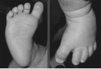
4.1.1.png

Clinical photograph of metatarsus adductus.
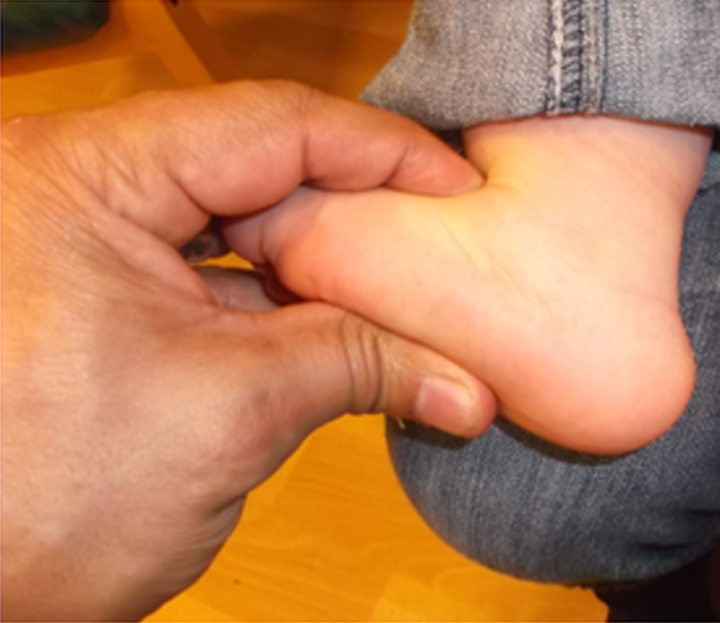
4.1.2.png

Hind foot is mobile and there is no equinus of the heel which is an important differentiating sign form clubfoot.
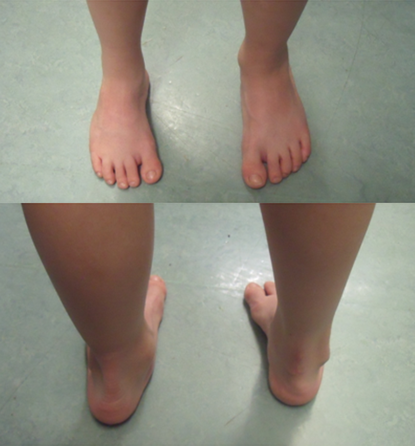
4.1.3.png

Metatarsus adductus can be part of residual club foot deformity (look for the foot size, hind foot deformity and any surgical scarring).

  • CTEV: Heel is normal in MA, Tendoachilles  (TA) is not tight in MA
  • Heel is in equinus and varus in CTEV, TA is tight
  • Skew foot: Hindfoot is in valgus in skewfoot
  • Internal tibial torsion (ITT): Foot thigh angle (FTA) and transmalleolar thigh angle (TMTA) is internal in ITT but heel bisector axis normal. Heel bisector axis is abnormal in MA but TMA is normal

4.1.4.png

This child was referred for Metatarsus adductus, however the lateral border is straight, heel bisector line passes between the second and the third digits but his FTA and TMTA are internal.

  • US to rule out DDH
  • X-ray in older child  to rule out skew foot
  • There is no need for X-ray just to confirm MA.
  • A number of radiological angles have been described to assess the severity of MA but their reproducibility as well as inter and intra rater reliability remains unproven.

4.1.5.png

This is a plain x-ray of a teenager with classical skew foot. Notice the valgus heel, abducted Midfoot and metatarsus adductus (Z-foot).

  • This is mostly a self-limiting condition.
  • Deformity that does persist does not cause a functional problem in the vast majority of cases.
  • Deformity might persist in around 15% of children or be slow to resolve.
  • Actively correctable deformity does not require any treatment.
  • Passively correctable deformity might benefit from stretching by the parents, although there is no clear evidence that stretching is effective.
  • Hindfoot is held in neutral and forefoot abducted with the other hand.
  • Fulcrum for abduction is over the lateral cuboid.
  • Serial casting may be an option especially for the more severe deformity.
  • Casting should be started early.
  • A recent randomized controlled trial has found that orthotic use gave clinically equivalent results to serial casting.

4.1.6.png

A child with MA being treated with serial casting. Notice the Pavlik Harness used for the co-existing dislocated hip.

  • Surgery is rarely required for rigid deformity not responsive to serial casting
  • It is difficult to estimate the true requirement for surgery in MA as some of the published series mostly included residual CTEV deformity rather than true MA
  • Indication for surgery would be persistent pain and/or footwear problem but this is rarely a childhood problem and mostly presents in adulthood.
  • A number of procedures have been described
  • These include:
    1. Abductor hallucis and 1st TMT release,
    2. Older children may benefit from opening wedge osteotomy of the medial cuneiform or metatarsal osteotomy.

There is controversy on the value of Medial metatarsocuneiform (MMC) and naviculcuneiform release.

  • Prognosis is generally excellent
  • Ponseti’s long term follow up series proved that these deformities can mostly be managed non operatively
  • Untreated MA is rarely problematic
  • Foot function is normal and degenerative arthritis does not develop.
  • It is important to manage parental concerns and reassure them
  • There are no long term reliable reports of outcome following surgery

There is also some evidence of an association between MA and later hallux valgus although it is not clear whether this is causative.

Previous
Next