TOPIC DETAILS

  Team Member Role(s) Profile
Paul Banaszkiewicz Paul Banaszkiewicz Section Editor
James Donaldson James Donaldson Segment Author

Rheumatoid arthritis

Definition

  • Rheumatoid arthritis (RA) is a chronic autoimmune inflammatory disease associated with pain,stiffness and swelling in the affected joints.
  • It is usually:
    1. Polyarticular – many joints are affected and these are classically symmetrical.
    2. Systemic – affects many other body systems.
    3. More common in women (as is characteristic of autoimmune diseases).
    4. Affects about 1% of the UK adult population.
    5. Peak onset in the fourth to fifth decade.
  • Unknown but thought to be multifactorial with environmental and genetic factors playing an important role.
  • HLA-DR4 genotype is particularly important.
  • Cell-mediated (T cell MHC type II) immune response against soft tissues.
  • Rheumatoid factor, an IgM antibody which recognises and attacks native IgG antibodies.
  • Immune complex is then deposited in end tissues such as the kidney as part of the pathophysiology.
  • Mononuclear cells are the primary cellular mediator of tissue destruction in RA. They phagocytose the IgM/IgG complexes along with fibrin and compliment. Necrosis of these cells induces release of lysosomal enzymes which causes an acute inflammatory reaction.
  • IL-1, TNF-alpha are part of a cascade that leads to joint damage.
  • Immune response is thought be related to infectious aetiology or HLA locus.
  • Antigen–antibody and antibody–antibody reactions occur.
  • Microvascular proliferation and obstruction.
  • Synovial pannus formation (histology shows prominent intimal hyperplasia).
  • Joint subluxation, chondrocyte death/joint destruction, and deformity.
  • Tendon tenosynovitis and rupture.

BS3RA 1.png

Fig 1. Immunopathogenesis of rheumatoid arthritis.

  • Inflammatory cells in the synovium results in progressive joint damage.
  • This classically causes painful, stiff, swollen joints especially in the morning (early morning stiffness).
  • Symptoms typically start gradually and progress over weeks to months.
  • Some have an acute onset of symptoms.
  • Symmetrical polyarthritis (commonest) – affects wrists, hands and feet, insidious low grade persistent inflammation.
  • Monoarthritis – usually one large joint affected, e.g. shoulder, knee.
  • Palindromic arthritis – bursts of severe monoarthritis moving rapidly from one joint to another that may settle after a few days.
  • Systemic disease – may be the presenting feature, e.g. heart disease (pericarditis), lung disease (pleural effusions, pleurisy, pulmonary fibrosis), weight loss, anaemia, fever.
  • Acute onset – widespread arthritis occurring in multiple joints.
  • Polymyalgic – limb girdle type pain and stiffness that progresses to joint synovitis.
  • A patient is said to have RA if he/she has satisfied at least four of the following criteria. Criteria 1–4 must have been present for at least 6 weeks:
  1. Morning stiffness in and around joints of at least 1 hour before improvement.
  2. Symmetrical joint swelling and involvement on both sides of the body.
  3. Arthritis of hand joints (at least one area swollen in a wrist, MTP or proximal interphalangeal (PIP)joint).
  4. Arthritis in at least three joint areas with swelling or fluid. The 14 possible joint areas are right/left PIP joints, metacarpophalangeal (MCP)joints, wrists, elbows, knees, ankles and MTP joints.
  5. Subcutaneous nodules over bony prominences or extensor surfaces.
  6. Radiographic changes typical of rheumatoid arthritis on PA hand and wrist radiographs that must include erosions or unequivocal bony decalcification localised to or most marked adjacent to the involved joints (osteoarthritic changes alone do not qualify).
  7. Positive rheumatoid factor.

Rheumatoid arthritis is a systemic disease, variably affecting many other systems as follows:

  • Cardiac – pericarditis, cardiomyopathy, nodules causing conduction or valvular defects.
  • Pulmonary – pulmonary fibrosis, pleural effusions, nodules (Figure 4) (granulomas), interstitial fibrosis, bronchiectasis
  • Eye – scleritis (Figure 2), episcleritis, Sjogren's syndrome (autoimmune condition affecting exocrine glands) dry eyes and dry mouth. Decreased secretions from salivary and tear duct glands.
  • Kidney – amyloid.
  • Nervous system – entrapment peripheral neuropathies.
  • Felty's syndrome (RA with splenomegaly and leukopenia) appears in patients with long-standing disease.
  • Rheumatoid vasculitis.
  • Still's disease (acute onset RA with fever, rash and splenomegaly).
  • Lymphoid tissue proliferation.

Always ask about activities of daily living in the history especially relating to fine movements of the hand (doing buttons, holding a pen) as these joints are commonly affected[V1] .
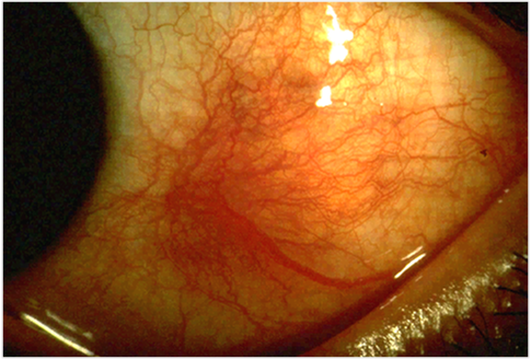
BS3RA 2.png

Figure 2. Scleritis  

BS3RA 3.png

Figs 3. Pulmonary nodules.

Signs on examination

  • Joint swelling (on palpation feels “boggy” due to synovitis), erythema, warm overlying skin, muscle wasting.
  • Synovitis mainly affects wrists, MCP, and PIP joints in the hand.

Classic RA hand and wrist signs (Figure 4)

  • Ulnar deviation of fingers
  • Wrist subluxation 
  • Boutonniere deformity (PIP flexion, DIP extension)
  • Swan-neck deformity (PIP extension, DIP flexion)
  • Z-deformity of thumb
  • Rupture of extensor tendons and muscle wasting
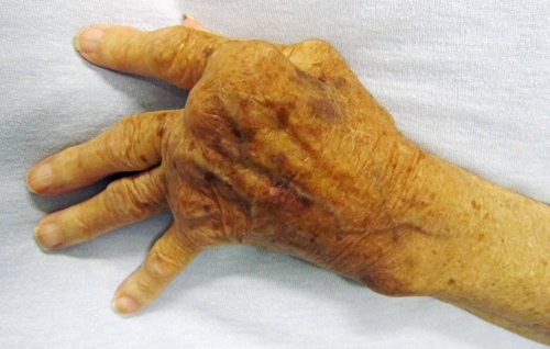
BS3RA 4.jpg

Figure 4. Rheumatoid wrist

Cervical spine

  • Cervical spine involvement is important as it can cause atlanto-axial joint (Figure 6) and subaxial subluxation (Figure 7).
  • Decreased space for spinal cord means that sudden movements of head or neck can cause cord damage and occasionally sudden death.
  • X-rays must be taken prior to general anaesthesia in RA patients so anaesthetist can assess this prior to intubation.
  • Other features on examination: rheumatoid nodules – swellings on elbows and fingers.
  • Carpal tunnel syndrome – nerve compressed due to synovitis.
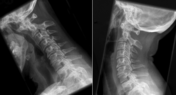
BS3RA 5.jpg

Figure 5. Cervical Myelopathy.Atlanto-axial subluxation in flexion.
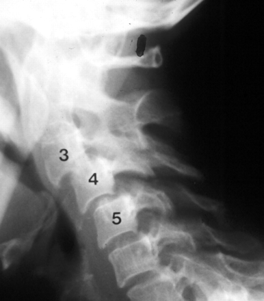
BS3RA 6.png

Figure 6. Rheumatoid Arthritis in the Cervical Spine. Subaxial subluxation C3-C5

Blood tests

  • FBC (anaemia) and inflammatory markers (ESR, CRP) useful for monitoring disease activity and treatment response.
  • Rheumatoid factor – found in 80% of RA patients, but often negative in early disease.
  • Not diagnostic for RA as 1% of general population will test positive.
  • Patients with rheumatoid nodules will always test positive.
  • Other blood tests are usually performed to exclude other causes of arthritis such as systemic lupus erythematosus.
  • Serum electrolytes, LFTs and bone profile (calcium and alkaline phosphate).
  • Immunological tests – antinuclear antibody (ANA) and autoantibody screen.
  • Rheumatoid factor may be positive in the presence of chronic infections (e.g. pulmonary tuberculosis), other rheumatological conditions (e.g. connective tissue disease) or sarcoidosis.

X-ray features (Figure 7)

  • Uniform narrowing of joint space 
  • Bilateral symmetrical joint involvement 
  • Periarticular soft tissue swelling 
  • Juxta-articular osteoporosis 
  • Large pseudocysts 
  • Bony erosions (late sign) 
  • Joint deformities – uniform narrowing of joint space 
  • Subluxation, carpal bone destruction
  • Magnetic resonance imaging may be helpful in diagnosing early synovitis
BS3RA 7.jpg
 
Figure 7. Widespread juxta-articular osteopenia and early erosion (irregularity of cortical surface). Carpal joints show loss of cartilage space and a mottled sclerosis of the carpal bones with discrete circular zones of deceased density consistent with cyst formation. There are numerous focal areas of cortical lysis.
  • This involves a multidisciplinary team approach.

Conservative

  • Regular exercise
  • Physiotherapy – exercises to improve joint range of motion and muscle strength
  • Occupational therapy – splinting of joints, appliances to assist activities of daily living

Medical

  • Regular analgesia – non-steroidal anti-inflammatory drugs (NSAIDs) provide symptomatic relief but do not affect RA progression.
  • Corticosteroids – injections or oral prednisolone (used early on in disease).
  • Disease-modifying antirheumatic drugs (DMARDs) can suppress disease activity and slow disease progression, e.g. methotrexate, gold, azathioprine.
  • Biological therapy:
  • Tumour necrosis factor alpha is a proinflammatory cytokine secreted by macrophages. It has a vital role in proinflammatory processes.
  • Anti-TNF therapies include infliximab, etanercept and adalimumab.
  • Biological therapies are delivered according to specific guidelines that require at least two other drugs to have been used first, one of which must be methotrexate.
  • The use of biological therapy is determined by NICE guidelines.

Surgical

  • Main options available in RA patients:
  • Arthroplasty – replacement of damaged joints 
  • Arthrodesis – fusion of smaller joints 
  • Synovectomy – removal of synovial lining of the diseased joint (usually done at wrist) 
  • Osteotomy – removal of bone to correct any deformity

BS3RA 8.png

Figure 8. Stages of arthritis

Gout

  • Is a disorder of nucleic acid metabolism causing hyperuricaemia and recurrent attacks of monosodium urate crystal deposition in joints.

Background

  • M:F = 20:1
  • Two types: 
  • Primary (95%): inherited disorder with overproduction or under excretion of uric acid
  • Secondary (5 per dent): myeloproliferative disorders, renal disease 
  • Only a small number of people with hyperuricaemia develop gout.

History

  • Galen (Figure 9) (129–199 AD), an ex-gladiatorial surgeon in the Pergamon arena in Asia Minor who moved to Rome, described gout as a discharge of the four humors of the body in unbalanced amounts into the joints (hence gout = gutta, a drop). 
  • Huber made the first radiological description of gout in 1896. 

BS3RA 9.jpg

Figure 9. Galen

Pathology

  • Humans lack the enzyme uricase, which is involved in elimination of excess nucleic acid purines and nitrogenous waste products through production and excretion of alantoic acid; hence in humans, uric acid is an end product of purines degradation (Figure 10).
  • Deposition of monosodium urate (MSU) crystals in synovial and periarticular tissue activate inflammatory mediators (Figure 11).

Figure 10. Hyperuricaemia and Gout. 

BS3RA 10.png

Figure 11. Gout and Hyperuricaemia Pharmacotherapy: A Pathophysiologic approach

Clinical

  • Recurrent arthritis
  • The joints most commonly affected by gout are: 
  • Those of the forefoot. Podagara– classic presentation of acute attack of first MTP joint (Figure 13).
  • The elbows and hands (Figure 14).
  • Unlike RA hand and wrist joints will have preserved joint spaces and normal mineralisation.
  • Large joints (hips, knees, ankles and shoulders) are infrequently involved.
  • The spine is very rarely affected. 
  • Associated with tophi (ear helix, eyelid, olecranon, Achilles tendon).
  • May be precipitated by chemotherapy for myeloproliferative disorders.
  • Nephrolithiasisis major extraarticular manifestation.
  • Pure uric acid stones are found in 80%, and uric acid is probably nidus for Ca-phos and oxalate calculi in remainder.

BS3RA 12.jpg

Figure 12. Gout 
 
BS3RA 13.jpg
 
Figure 13. Gout 

Radiology

  • The radiographic changes occur late in the course of gout, usually associated with the chronic tophaceous stage. 
  • Joints show signs of a degenerative arthritis with joint space narrowing, osteophyte formation and sclerosis (Figure 15). 
  • Later, the joint may be become deformed or ankylosed.
  • Well circumscribed, “punched out” cystic lesions may be seen adjacent to the cortex of an affected joint (Figure 16). 
  • Cysts larger than 5 mm are suggestive of gout, cf. OA.
  • Tophaceous material may eventually breech the cortex with extensive bone destruction.
BS3RA 14.png
Figure 14. Gout 
 
BS3RA 15.png
Figure 15. Gout 

Laboratory

  • Hyperuricemia
  • Biochemical hallmark of gout, but not by itself diagnostic
  • Leukocytosis 
  • Increased ESR 
  • Synovial fluid 
  • Leukocyte counts = septic arthritis 
  • Viscosity is less than that seen in a septic or inflammatory arthritis 
  • MSU needle-like intracellular and extracellular crystals 
  • Negatively birefringent crystals under polarised light microscopy are diagnostic for gout (Figure 17)
BS3RA 16.jpg
Figure 16. MSU crystals viewed under polarized light with a red plate that makes those in the plane of the long axis of the red plate yellow, which indicates that they are negatively birefringent.

Treatment

  • Acute
  • Indomethacin
  • Colchicine: inhibits the inflammatory mediators
  • Chronic
  • Allopurinol for hyperuricaemia and tophi 
  • Acute arthritis caused by calcium pyrophosphate dihydrate (CPPD) crystal-induced inflammation 
  • May perfectly mimic gout during acute flare 
  • Attacks occurring before age 50 years are uncommon 

Clinical

  • Most often affects the knee and the wrists 

Radiology

  • Calcification densities in hyaline or fibrocartilage, which are found in knee menisci (Figure 18), acetabular labrum and triangular fibrocartilage complex (TFCC).
BS3RA 17.gif

Figure 17. Chondrocalcinosis knee

Laboratory

  • CPPD crystals are visualised under compensated .
  • Crystals may be more difficult to detect than MSU crystals because of their smaller size, more intralysosomal location, and less brilliant colours.
  • CPPD crystals show weak positive birefringency and have squared or rhomboidal shaped ends (Figure 19).
  • Alizarin red stain can confirm that these clumps are masses of calcium crystals.
BS3RA 18.png
Figure 18.  CPPD crystal viewed under polarized light with a red plate. The crystal is aligned in the long axis of the red plate, so that it is bluish-white, which indicates that it is weakly positively birefringent.

Treatment

  • Aspiration of the involved joint and steroid injection, once diagnosis of infection has been excluded, will usually control symptoms.

Systemic lupus erythematosus

  • Is a chronic inflammatory disease.
  • Usually affects women (especially Afro-Caribbean descent).

Clinical features include:

  • Fever
  • Butterfly malar rash (Figure 20)
  • Pancytopenia
  • Pericarditis
  • Nephritis
  • Polyarthritis
  • The commonest feature is joint involvement (75%)
  • Arthritis causes acute, red and swollen joints, often the PIPJs, MCPJs, carpus and knees
  • It is not as destructive as RA
  • Mortality is generally from renal disease
  • ANA+ve, HLA-DR3, plus or minus RF+ve
BS3RA 19.png
Figure 19. SLE malar rash
  • Common among the elderly
  • Aching and stiffness of shoulder and pelvic girdle
  • Associated with malaise, headaches and anorexia
  • Raised ESR, anaemia, raised ALP, increased immune complexes
  • Treated with steroids
  • May be associated with temporal arteritis

Three major types:

    • Systemic (20%)
    • Polyarticular (50%). More than five joints involved
    • Pauciarticular (30%). Less than four joints involved
  • Sero+ve juvenile rheumatoid arthritis (JRA) is associated with chronic, active and progressive disease.
  • Early onset (before teens) or late onset (teenager or later).
  • Sero+ve polyarticular JRA often develops into adult RA with joint destruction.
  • Pauciarticular JRA is associated with iridiocyclitis in 50% of cases.
  • Multiple HLA associations including DR2, DR4, DR5, DR8 and B27 in boys.
  • HLA-B27 (chromosome 6, D locus) +ve and –ve RF titre
  • Bilateral sacroilitis with or without acute anterior uveitis.
  • Insidious onset of back pain and associated morning stiffness, as well as hip pain in third to fourth decade.
  • Progresses over 20 years often with progressive spinal flexion deformities (Figure 21).
  • Male/female 3 to 10:1.
BS3RA 20.png
Figure 20. Ankylosing spondylitis 

Radiographic features

  • Squaring of vertebrae
  • Vertical syndesmophytes “bamboo spine”
  • Obliteration of SI joints
  • “Whiskering” of enthesis
  • Diffuse osteoporosis
  • Ascending ankylosis usually begins in the T-L spine (Figure 22).
  • Chin on chest deformity requires corrective osteotomy of cervicothoracic junction.
  • Cervical spine fractures are common. Associated with epidural haemorrhage and high mortality; 75% will develop neurology.
  • Severe kyphotic deformities require posterior closing wedge osteotomies.
  • Associated with protrusio acetabuli, heart disease and pulmonary fibrosis.

Extra-skeletal manifestations include:

  • Iritis
  • Aortitis
  • Colitis
  • Arachnoiditis
  • Renal amyloidosis
  • Sarcoidosis
  • Pulmonary fibrosis
  • Poor prognosis if pulmonary involvement, hip involvement and early age of disease onset.
BS3RA 21.jpg
Figure 21. Ankylosing spondylitis.Bamboo spine

Triad of: 

    • Conjunctivitis
    • Urethritis
    • Oligoarticular arthritis
  • Can’t see, pee or bend the knee!
  • Recurrence is common
  • 80–90% are B27+ve, 60% have sacroilitis
  • Affects 5–10% of patients with psoriasis
  • B27+ve in 50%
  • Many forms
  • Commonly oligoarticular and assymetrical affecting the small joints of the hand (Figure 23) and feet

Features:

  • Nail pitting, fragmentation and discolouration
  • Sausage digits
  • Pencil in cup deformity with DIP involvement
BS3RA 22.jpg
Figure 22. Psoriatic arthropathy.Clinical picture hands

BS3RA 23.jpg
Figure 23. Psoriatic arthropathy.Radiographs hand.Erosive changes 
  • 10–20% of Crohn’s disease and ulcerative colitis patients develop arthritis, commonly the large weight-bearing joints.
  • 5% experience axial disease.
  • Non-deforming, usually acute monoarticular synovitis may preceed bowel symptoms.
  • B27+ve in 50%; associated with ankylosing spondylitis in 10–15%.
Previous
Next