TOPIC DETAILS

  Team Member Role(s) Profile
Fazal Ali Fazal Ali Section Editor
Mark Malek Racy Segment Author

Wrist and Hand history 

Main demographic details

  • Age
  • Occupation
  • Handedness
  • Hobbies/ recreational activities

Nature of complaint

Pain

  • Constant/ intermittent?
  • Sharp/ dull
  • Burning/ shooting
  • Exacerbating/ relieving factors
    • Specific actions e.g. opening a jar

Swelling

  • Painful?
  • Variable size?

Stiffness

  • Which movements are affected?
  • Is this limiting function?

Weakness

  • Due to pain?
  • When do they notice the weakness?

Onset

  • Insidious
  • Chronic

Associated symptoms

  • Clicking
  • Clunking

Effect on ADLs/ work/ hobbies

Basics

Sit opposite the patient, ensure adequate exposure (above elbows) and examine both hands side by side.

The examination begins with a screening test of look, move, feel and then based on the identified pathology or site of tenderness, specific tests are carried out

Look

Put the hands next to each other in a similar position and start inspection on the dorsum of the wrist. Look for obvious swellings, deformities or scars. Is the deformity unilateral or bilateral?

Ask the patient to supinate and assess the volar wrist.

Finally ask the patient to flex their elbows and show you the ulnar aspect of their wrists and the extensor surface of their forearms.

Deformities to look for:

Scars

Rheumatoid arthritis

  • Bilateral with multiple joint involvement
  • Diffuse dorsal swelling/ synovitis
  • DRUJ subluxation
    • Dorsal prominence of the ulnar head
    • Asymmetry when assessing the ulnar border of the wrists with elbows flexed
  • Ulnar and volar subluxation of the carpus
  • Other findings to note:
      • Radial metacarpal drift/ ulnar deviation of the fingers
      • Extensor tendon rupture/ Vaughan-Jackson syndrome
      • Flexor tendon rupture
      • Finger deformities
      • Rheumatoid nodules

Radial malunion

  • Radial shortening
  • Prominent ulna

Madelung’s deformity

  • Defective development of the volar/ulnar distal radial physis
  • Volar radial bowing with a dorsally prominent distal ulna
  • Can be bilateral in up to 60%
  • Called a ‘reverse Madelung’s deformity’ where  there is dorsal radial bowing

Wrist ganglion

  • Most commonly dorsal, overlying the scapholunate ligament

Carpal boss

    • (normally) painless bony prominence at the base of the second and third metacarpals
    • Tends to be more distal than dorsal ganglia

Muscle wasting
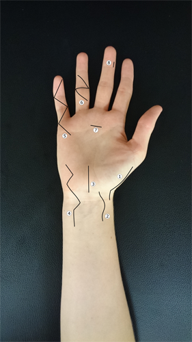
Scars volar copy.png

Figure 1.Volar scars 1.Wagner(a)Trapeziectomy (b) Bennett fracture 2 Scaphoid 3 Carpal tunnel 4 Guyon’s canal 5 Brunner (a)Dupuytren’s (b)Flexor tendon repair 6 Z-plasty (a)Scarring (b)Dupuytren’ 7 Trigger finger release 8 Combined with (7): flexor tendon sheath washout
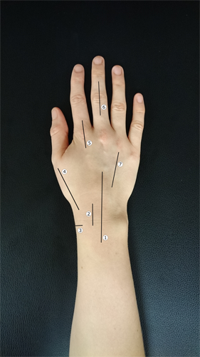
Scars dorsal.png

Figure 2.Dorsal scars 1.Midline dorsal (a)Fusion (b)Synovectomy (c)Carpal ligament repair (d)1+3+4: EIP to EPL transfer 2 Dorsal to scaphoid (a)Proximal pole fractures (b)Ligament repair 3 De Quervain’s release 4 Radiopalmar to thumb base (a)Trapeziectomy (b)Bennett fracture 5 Dorsal to MCPJ 6 Dorsal to IPJ (a)Tendon repair (b)Arthroplasty (5)Dorsal to metacarpal (a)Fracture fixation

Move

Each movement can be assessed as part of a screening test or in more detail using a goniometer, with the radius and middle finger metacarpal as references.

Flexion/ extension

  • Dorsiflexion- place the palms together and raise the elbows to position the forearms horizontal. A loss in range will show as one elbow being lower than the other.
  • Palmarflexion- as above but with the dorsum of the hands in contact, bringing the forearms into the horizontal plane

Normal values:

    • Dorsiflexion:     75°
    • Palmarflexion:   75°

Dorsiflexion.JPG

Figure 3.Wrist dorsiflexion

Palmarflexion.JPG

Figure 4. Wrist palmarflexion

Pronation/ supination

  • The patient should make a fist, holding a pen in each hand.
  • With elbows tucked in to their side and flexed to 90 degrees, assess pronation and supination
  • Holding thumbs out instead is an option (see photo), but patients can appear to have increased range of movement by moving their thumbs

Normal values:

    • Pronation:        75°
    • Supination:       80°

Pronation.JPG

Figure 5.Wrist pronation

Supination.JPG

Figure 6.Wrist supination

Radial/ ulnar deviation

  • Assessed with the forearms pronated and elbows tucked into the patient’s sides.

Normal values:

  • Radial deviation:           20°
  • Ulnar deviation:            35°

Goniometer wrist flexion.JPG

Figure 7. Measuring wrist flexion with a goniometer

Feel

Start radial/ dorsal and work round ulnar/ volar.

As the wrist is a superficial joint the site of tenderness guides to the underlying structure involved and therefore which provocative test is to be used.

Surface anatomy volar numbered.png

Figure 8 .Dorsal surface anatomy

  1. Lister’s tubercle
  2. Radial styloid
  3. 1st extensor compartment/ distal radius
  4. Anatomic snuffbox
  5. 1st CMC joint
  6. Scapholunate ligament
  7. Lunotriquetral ligament
  8. DRUJ
  9. TFCC
  10. Ulnar styloid

Surface anatomy volar numbered.png

Figure 9.Volar surface anatomy

  1. Scaphoid tubercle
  2. Radial artery
  3. Pisiform
  4. Hook of hamate

Provocative tests

These tests are performed once you have identified a particular pathology from your screening examination. Depending on the location of pain, tenderness or deformity, the relevant provocative or instability tests are performed.

Where there is more than one test for a particular pathology, the most important one is highlighted in bold (e.g. Kirk-Watson test for scapholunate instability).

When performing the various tests, ensure that the tested movements are occurring at the relevant joint (i.e. the wrist) instead of the hand.

Compare all tests to the opposite side.

De Quervain’s tenosynovitis

This test for De Quervain’s tenosynovitis aims to stretch the tendons of the first extensor compartment; the abductor pollicis longus and extensor pollicis brevis tendons.

Finkelstein’s test

  • Adduct the thumb/ place it inside a fist and ulnarly deviate the wrist
  • Positive result: reproduction of pain

Scapholunate instability

Scapholunate ligament injuries are often missed and demonstrate a spectrum of severity. The amount of displacement or laxity should be compared to the opposite side and can represent the extent of instability.

Key points from the history:

  • Wrist loaded in extension, ulnar deviation and intercarpal supination (e.g. falling on pronated hand)
  • Often high energy injury
  • Pain on loading dorsiflexed wrist
  • Reduced grip strength, clicking or pain

Scaphoid shift/ Kirk Watson

  • Flex the patient’s elbow so the forearm is vertical
  • Use one hand to hold the patient’s wrist
  • Your thumb should rest on and put pressure on the scaphoid tubercle, with the index finger over the scapholunate interval dorsally
  • Whilst maintaining dorsally directed pressure on the scaphoid tubercle, move the wrist from ulnar to radial deviation with your other hand
  • As the wrist moves into radial deviation, the scaphoid should flex and feel more prominent on the examiner’s thumb
  • With a scapholunate ligament injury, however, the scaphoid subluxes dorsally as the wrist moves into radial deviation, felt as a click or pressure on the index finger
  • Can be normal in up to 36%

Scaphoid thrust

  • The hand is held in the same way as the scaphoid shift test
  • The wrist is gently cycled between radial and ulnar deviation to relax the patient
  • With the wrist in slight radial deviation, press with your thumb on the scaphoid tubercle
  • Positive test: the scaphoid is felt moving dorsally

Scaphoid lift

  • Pronate the wrist
  • With one hand, stabilise the lunate between your thumb volarly and index finger dorsally
  • With the opposite hand, hold the scaphoid in the same manner and lift the scaphoid dorsally (and volarly) for a positive test


Video 2.Scaphoid shift/ Kirk Watson test for scapholunate instability.

Lunotriquetral instability

Caused by injuries to the lunotriquetral ligament

Key points from the history:

  • Wrist loaded in forced dorsiflexion +/- radial deviation
  • Ulnar sided pain, worse on power grip

Ballotment (Reagan)

  • With one hand, stabilise the lunate between your thumb and index finger on the volar and dorsal sides respectively
  • With the opposite hand, hold the triquetrum (and pisiform) and attempt to shift them in dorsal and volar directions
  • Positive result: laxity, crepitus or pain produce a positive result

Shear (Kleinman)

  • With your contralateral hand, place your thumb over the lunate dorsally
  • Place your index finger over the triquetrum/ pisiform on the volar surface
  • Stabilising the lunate with your thumb, press on your index finger, displacing the triquetrum dorsally

Midcarpal instability

From radial to ulnar deviation, the proximal carpal row moves from flexion to extension, with smooth translation of the distal row from volar to dorsal. Where there is instability between the two rows, this combined movement is disrupted and a ‘clunk’ is often felt as the two rows realign.

Key points from the history:

  • High energy dorsiflexion injury
  • Radial styloid fracture
  • Clicking and pain when lifting heavy objects
  • Reduced grip strength

Midcarpal shift (Lichtman)

  • With one hand, grasp the patient’s hand with your thumb lying over the capitate dorsally
  • Support the forearm with the opposite hand
  • Start by applying a volar directed force with your thumb on the capitate and note the amount of translation (graded I-IV)
  • Apply an axial force to the carpus and move the wrist from full radial to ulnar deviation
  • Positive result: painful clunk


Video 3.Midcarpal shift/ Lichtman’s test for midcarpal instability.

Ulnar sided pathology

There are a range of causes for ulnar sided wrist pain, including those listed below. Often they are interlinked, such as with TFCC tears and DRUJ instability. The ulnocarpal stress test can be used as a general screening tool to confirm pathology in this area.

Common causes:

  • TFCC tears
  • DRUJ instability
  • Lunotriquetral injuries (described earlier)
  • Pisotriquetral OA
  • Hamate fracture
  • ECU subluxation

Ulnocarpal stress

  • Support the patient’s hand with your contralateral hand and grasp the forearm in your other hand
  • Ulnarly deviate the wrist and apply axial compression
  • Slowly dorsiflex and palmarflex the wrist
  • Alternatively, keep the hand supported and rotate the forearm
  • Positive result: ulnar sided wrist pain

Triangular fibrocartilage complex

Injuries to the TFCC can be degenerative or traumatic and associated with DRUJ injuries.

Key points from the history:

  • Rotational injuries or fall on pronated dorsiflexed wrist
  • Supination (e.g. using a door key) causes ulnar sided wrist pain

Fovea sign

  • Palpate the ulnar border of the wrist just distal to the ulnar styloid, dorsal to the FCU tendon
  • Positive result: tenderness

TFCC compression test

  • Pronate the wrist fully and support the forearm with one hand and the hand with the other
  • Axially load the wrist in ulnar deviation
  • In this position, pronate and supinate the wrist
  • Positive result: ulnar sided pain

Fovea sign.png

Figure 10.Fovea sign for TFCC injuries

U: Ulna

T: Triquetrum

H: Hamate

P: Pisiform

M: 5th Metacarpal

FCU: Flexor carpi ulnaris tendon

ECU: Extensor carpi ulnaris tendon

*: Fovea/ site of tenderness

DRUJ

Pathology to the distal radioulnar joint can be post traumatic or degenerative, from osteoarthritis or inflammatory arthritis. Pain from the compression test can suggest injury or degeneration whereas the other tests identify acute or chronic subluxation.

Key points from the history:

  • History of radius/ ulna fracture
  • History of inflammatory arthropathy
  • Pain on pronation/ supination
  • Reduced grip strength

Piano key

  • Support the patient’s wrist with your ipsilateral hand
  • Using your other hand, press down on the dorsal distal ulna
  • Positive result: increased motion (more than 5mm to opposite side) and ulna should spring back like a piano key when released

Compression

  • Support the patient’s hand with your contralateral hand
  • With your other hand, squeeze the distal radius and ulna together and using the same hand, pronate and supinate the forearm
  • Positive result: pain

Radioulnar drawer

  • With one hand, hold the radius between your thumb and index finger
  • With your opposite hand, hold the ulna in the same manner and attempt to move the ulna in dorsal and volar directions
  • Positive result: laxity with or without pain

Dimple sign

  • Grasp the forearm with one hand, applying a volarly directed force to the dorsal surface of the distal ulna
  • Apply traction across the wrist joint
Previous
Next