TOPIC DETAILS

No contributor information available.
Previous
Next

QUESTION 1 OF 10

A 26-year-old falls and injures her right hand. There is no bony injury identified on x-ray, but she is noted to have a lytic lesion at the base of her middle finger proximal phalanx. The lesion demonstrates geographic bone destruction, bony expansion and cortical thinning. The lesion is asymptomatic.
What is the most appropriate management plan for this patient?

QUESTION ID: 1254

1. CT Scan
2. MRI scan and review
3. Reassurance and discharge
4. Reassurance and observation with serial radiographs
5. Screening Blood tests and chest x-ray

QUESTION 2 OF 10

54.A 9-year-old boy is brought to ED by his stepfather. He complains of right arm pain but does not give a clear history of fall. X-rays show as below
Which of the following is the most likely diagnosis?

ks 6.png

QUESTION ID: 1277

1. Aneurysmal bone cyst
2. Fibrous dysplasia
3. Giant cell tumour
4. Non-accidental injury
5. Unicameral bone cyst

QUESTION 3 OF 10

56.A 30-year-old female presents to the ED with swelling in the back of knee and difficulty in bending the knee. Radiographs  demonstrate  a large lobulated mass attached to the posterior cortex of the femur.

KS8 .png
Which of the following statements is NOT TRUE about this tumour?

QUESTION ID: 1280

1. An X;18 translocation is characteristic
2. De-differentiation into a higher-grade tumour is infrequent but possible
3. It is associated with an excellent prognosis, with over 90% survival at 5 years
4. Radiotherapy before surgery is not required
5. Treatment is complete surgical excision

QUESTION 4 OF 10

A 30-year-old female physiotherapist presents with left wrist pain and swelling with no history of trauma. X-ray demonstrates an eccentric, lytic lesion in the distal radius. MRI demonstrates a breach in the cortex with a small soft tissue component. Biopsy reveals a tumour comprising multinucleated giant cells and stromal cells. female physiotherapist presents with right wrist pain and swelling with no history of trauma. X-ray demonstrates an eccentric, lytic lesion in the distal radius. MRI demonstrates a breach in the cortex with a small soft tissue component. Biopsy reveals a tumour comprising multinucleated giant cells and stromal cells.
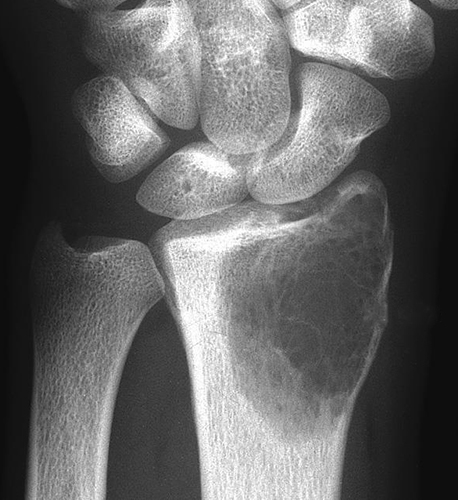
KS 10.jpg

Which of the following statements is NOT TRUE about this tumour?

QUESTION ID: 1281

1. Campanacci system is used for radiological grading
2. Denosumab is a recognised adjuvant treatment in selected cases
3. Patients with lung metastases have a poor outcome
4. Recurrence rates can be high following curettage
5. This tumour rarely affects skeletally immature patients

QUESTION 5 OF 10

A 15-year-old girl has a painful thoracolumbar scoliosis, worse at night and relieved by Ibuprofen. 
What is her most appropriate definitive treatment?

QUESTION ID: 1315

1. Adjuvant chemotherapy and surgical excision
2. Adjuvant radiotherapy and surgical excision
3. CT-guided radiofrequency ablation
4. En block surgical excision
5. Radiotherapy alone

QUESTION 6 OF 10

115.A 16-year-old female presented with a painful hand after punching a wall, her x rays are showed below.
Which of the following is not correct:

12. A 16-year-old female presented .png

QUESTION ID: 2259

1. A bone scan will show increased uptake
2. Association with vascular malformations in the hand suggests a risk malignant transformation of 60-80%
3. In patients with the underlying pathology the radiological features of cortical thinning and expansion is present in the hands and feet but not seen in the long bones
4. The appearance in a long bone can mimic bone infarct areas.
5. The pathology is due to failure if endo-chondral ossification

QUESTION 7 OF 10

116.Looking at the following x rays which is the most appropriate description:

Figure 1 X ray 1.jpg

Figure 1 X-ray 1

Figure 2 X ray 2 .jpg

Figure 2 X-ray 2

Figure 3 X ray 3.png
QUESTION ID: 2260

1. The calcified fragment seen within the lucent area on x ray 1 is suggestive of an early healing of an intramedullary fracture
2. X ray 1 likely to be an active unicameral bone cyst
3. X ray 2 is likely to be a latent aneurysmal bone cyst
4. X ray 2 is likely to be an aneurysmal bone cyst as it extending to the physis
5. X ray 3 is likely to be an active unicameral cyst as it’s not wider than the physis

QUESTION 8 OF 10

A 10-year-old boy presents with a painful swelling in the index finger of the right hand. This has grown slowly over 6 months. Plain x-rays show multiple osteolytic lesions in the small bones of the hand, with expansion of the middle phalanx of the index finger.
Which of the following statements is true?

QUESTION ID: 2261

1. Maffucci syndrome presents with multiple osteochondromas and haemangiomas
2. Ollier disease presents with multiple osteochondromas and haemangiomas
3. Ollier disease shows autosomal dominant Inheritance
4. Persistent pain and growth of an enchondroma raises the possibility of malignant transformation
5. The risk of malignant transformation in Ollier disease is close to 100%

QUESTION 9 OF 10

11.A 25 year old footballer presents with 2 months of pain and swelling in his ring finger. The finger is causing paroxysmal pain with cold intolerance and tender to touch. There is no clear history of previous injury on the finger. 
Examination reveals a small bluish nodule near the nail bed

ks 5.jpg

QUESTION ID: 2265

1. Radiotherapy
2. Curettage and bone grafting
3. Marginal excision
4. Prolonged course of antibiotics
5. Sclerotherapy

QUESTION 10 OF 10

15. A 10-year-old boy presents with a painful swelling in the index finger of the right hand. This has grown slowly over 6 months. Plain x-rays show multiple osteolytic lesions in the small bones of the hand, with expansion of the middle phalanx of the index finger.
Which of the following statements is true?


QUESTION ID: 2276

1. Maffucci syndrome presents with multiple osteochondromas and haemangiomas
2. Ollier disease presents with multiple osteochondromas and haemangiomas
3. Ollier disease shows autosomal dominant Inheritance
4. Persistent pain and growth of an enchondroma raises the possibility of malignant transformation
5. The risk of malignant transformation in Ollier disease is close to 100%