TOPIC DETAILS

  Team Member Role(s) Profile
SattAR Sattar Alshriyda Section Editor
  • CTEV is a common congenital limb anomaly.
  • Wynne Davies in her classic paper estimated incidence in Devonshire to be 1 in 1000 live births that is widely considered to be the standard UK incidence.

4.2.1.png

Pre-natal diagnosis of CTEV using 3DUS, Obstetric Imaging Martin SR, Cedar JM. © Saunders (used under licence).

  • Etiology of CTEV is unclear.
  • A number of genes have been identified and it is now felt that   inheritance is likely to follow a multifactorial threshold model.
  • Results from the Danish Twin registry suggest a 33% concordance rate in a monozygotic twin.
  • First degree relatives are at increased risk compared to the general population. 
  • In most of the cases the cause for CTEV is unknown and it is labeled as “idiopathic”
  • Associated with other congenital anomalies in 20% cases.
  • This includes Spina bifida, Arthrogryposis,Cerebral palsy and other neurological disorders, chromosomal defects, generalized systemic abnormalities.
  • The association with DDH is controversial. Both increased and decreased risk has been identified. Results from a recent UK study would suggest that idiopathic CTEV may not be a strong risk factor for pathologic (Graf Type III) type of DDH

4.2.2.png

Specimen from a 20 wk old fetus. Fukuhara K, Schollmeier G, Uhthoff HK. The Pathogenesis of Clubfoot. J Bone Joint surg (Br) 1994. 

  • The foot goes through a number of development phases in utero, eventually attaining the normal shape at the final foetal phase.
  • There has been suggestion that interruption of growth at the foetal stage might result in clubfoot deformity.
  • Intra-uterine CTEV is not detected until the 16th week of pregnancy.

4.2.3.png

From Fukuhara K, Schollmeier G, Uhthoff HK. The Pathogenesis of Clubfoot. J Bone Joint surg (Br) 1994.

The top image is from a foetus with severe CTEV and the bottom image is from a normal foot. Compare the difference in type III collagen staining between the two.

  • Researchers have found increased  collagen fibres and myofibroblast like cells in the medial soft tissue structures of the foot in CTEV patients.
  • This suggests that a fibromatosis type process might be involved in CTEV.
  • Another common finding is absence or hypoplasia of the anterior tibial and dorsalis pedis arteries.
  • This may indicate arrested development of the leg before the final foetal phase.

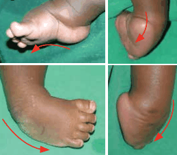
The anatomical abonormalities are: C: cavus of the midfoot, A: adduction of the midfoot, V: varus of the hindfoot and E: equinus of the hindfoot.

  • Forefoot: great toe is plantar-flexed, the forefoot is pronated relative to the hindfoot
  • Midfoot: midfoot is adducted at the talo-navicular joint.
  • Both talus and navicular are medially displaced, inverted and navicular is plantarly subluxed on the talar head.
  • Talar head is uncovered laterally and clinically palpable.
  • Talar neck is short and medially deviated and talus is in equinus.
  • Cuboid is also medially displaced on the calcaneo-cuboid joint.
  • Hindfoot: hindfoot is is equinus and varus due to plantar-flexion and inversion of the calcaneus.
  • Ankle: the tibio-talar joint is normal.

Medial soft tissue structures are short, thickened and medially displaced. Therefore they act both as tethering and deforming forces.

4.2.4.png

Relationship of Calcaneus ( green), talus (red) and navicular (yellow):  the normal foot on the left and CTEV on the right.

4.2.5.png

MRI image of gradual change in TNJ and CCJ before ( left) and after treatment (right). Navicular (red), Talus (blue), Calcaneus (yellow), cuboid (green).

From Staheli L. Clubfoot: Ponseti management. © Global help.

  • When a neonate presents in the clinic with CTEV a complete head to toe examination is essential to rule out other congenital abnormalities.
  • The spine and the joints should be specifically examined to rule out spina bifida and arthrogryposis.
  • Differential diagnoses include metatarsus adductus ( MA) and positional clubfoot ( a foot that is fully correctable on gentle handling).
  • Apart from the foot the calf muscle usually has reduced girth and length and the foot may be smaller than the contralateral side.
  • Hindfoot is the key for examination and is normal in both MA and positional clubfoot but fixed and abnormal in CTEV ( and CVT). However, both MA and positional clubfoot may be associated with intra-uterine moulding and are known risk factors for DDH.

4.2.6.png

Bilateral CTEV.

4.2.7.png

Typical deformities of CTEV. Clockwise from left: cavus, adductus, equinus, varus. From Staheli L. Clubfoot: Ponseti management. © Global help.
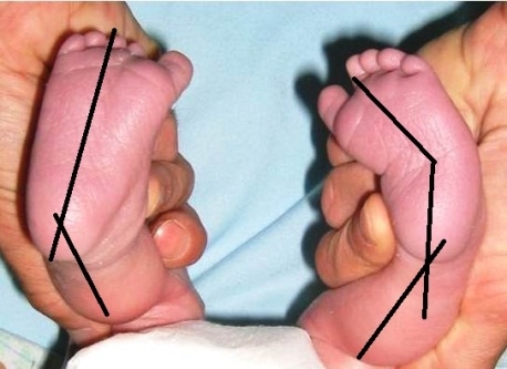
4.2.8.jpg

Unilateral CTEV, compared to the normal side the forefoot is adducted and the hindfoot is in varus.
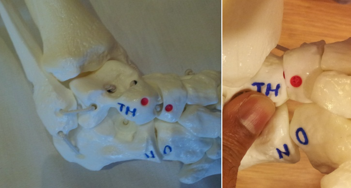
4.2.9.png

Model showing an uncorrected CTEV (left); talar head is prominent and palpable in front of lateral malleolus. Anterior calcaneal process is not palpable. Corrected CTEV (right); anterior process of calcaneum is now palpable as STJ is reduced.


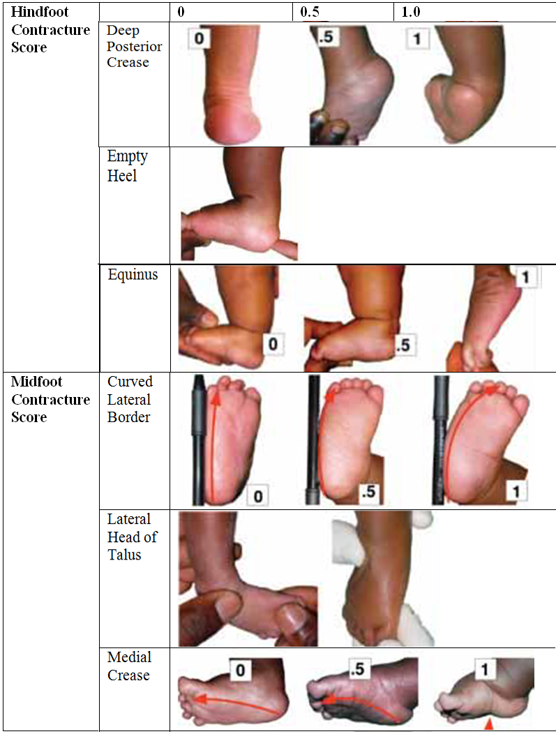
Pirani scoring:

  • This is a clinical grading system of the severity of deformity.
  • It does not take into account the functional state, radiological status or the gait.
  • The score has been widely validated.
  •  It can be used to monitor the progress of treatment and is also predictive of the number of casts required.
  • The assessment is divided into midfoot   (3parts) and hindfoot (3 parts). Each parameter can have a score of 0, 0.5 or 1. The more severe the foot the higher the score.
  • The foot and ankle should be placed in maximal correction for assessment. A sequence of look, feel, move helps to commit the steps to memory: 

Hindfoot score: 0-3

Look: posterior heel crease:

Multiple fine creases: 0

One or two deep creases: 0.5

Deep creases change the contour of the arch : 1

Feel: empty heel sign:

Calcaneal tuberosity easily palpable : 0

Calcaneal tuberosity difficult to palpate : 0.5

Calcneal tuberosity not palpable: 1

Move: equinus:

Full ankle dorsiflexion : 0

Ankle in plantigrade: 0.5

Ankle in equinus: 1


Midfoot score: 0-3

Look: curvature of lateral border: ( place a pencil against the lateral border)

Straight border: 0

Mild, distal curved border: 0.5

Lateral border curves at CCJ: 1

Feel: talar head:

Talo-navicular completely reduced, talar head not felt : 0

Talo-navicular joint partially reduced, talar head difficult to feel: 0.5

Talo-navicular joint unreduced, talar head easily felt: 1

Move: medial crease: (assessed after moving the foot to corrected position)

Multiple fine creases: 0

One or two deep creases: 0.5

Deep creases change the contour of the arch: 1

  • DiMeglio has proposed a 20 point classification system. Both of the scoring systems have good inter-rater reliability but none predict long term outcome.
  • Most of the delegates attending a recent European consensus meeting on Clubfoot felt that the Pirani scoring method was simpler to use and favoured it over the DiMeglio scoring method.
4.2.10.png

Pirani scoring

Depending on severity CTEV can be classified as:

  • Postural: moulding defect, easily correctable
  • Idiopathic: typical CTEV
  • Complex/atypical CTEV: very tight hindfoot and plantar structures
  • Syndromic: associated with other congenital bonormalities

CTEV can also be classified depending the presentation:

  • Early: typical ante-natal presentation
  • Delayed/neglected: presents anytime after walking age
  • Resistant: deformity not amenable to Ponseti regime
  • Relapsed: recurrence of deformity after initial correction

US scan: although the majority of patients are diagnosed at birth CTEV can be reliably diagnosed pre-natally at 18-20 week scan. It has high sensitivity and specificity. It is recommended to arrange appropriate counselling for parents following pre-natal diagnosis.

Although there is controversy regarding the association with DDH we prefer to perform hip US scan in all new patients to rule out DDH.

Xray: xray is not  necessary for initial management. Most of the newborn’s foot is cartilaginous. However the talo-calcaneal (TCN) angle is abnormal in CTEV. Normal TCN configuration in both the AP and lateral view is that of an open scissor but in CTEV the TCN appearance is that of a closed scissor with parallelism of talus and calcaneus.

Xray would also be of use to confirm ossification of lateral cuneiform and size of ossified bone in young patients if decision is made to transfer the Tibialis Anterior (ATT) tendon.

Goal of CTEV management is to achieve a pain-free shoeable foot with normal functional ability. However there is no clear agreement regarding the parameters for successful outcome. Parents often worry if their child would be able to undertake sports.

Ponseti’s research has revolutionised the management of CTEV and 95% of patients can be managed successfully with his technique. A 30 year follow up study from Ponseti’s original centre conducted by Copper and Dietz found that 78%of patients who had successful Ponseti treatment reported good or excellent functional outcome compared to 85% of normal participants with no foot deformity. Dobbs’ long term follow up of clubfeet with extensive soft tissue surgery on the other hand found that patients had significant functional impairment that appeared to directly correlate with the extent of soft tissue surgery.

The Ponseti technique comprises of:

  1. Gentle manipulation
  2. Serial casting
  3. Achilles tendon tenotomy in 90%
  4. Maintenance of correction with a regime of splinting with the foot abduction brace

Each component requires the same careful attention to detail to achieve good results.

4.2.11.png

Ponseti technique: The first ray is elevated and soft tissues gently stretched. Attention to the details is vital for successful outcome. Image 2 showed Inadequate correction: 1st ray is still plantar-flexed. Image 3&4 showed adequate elevation of first ray and to ensure that the cavus component is fully corrected.

  • Ponseti casting: casting should be started within the first 4 weeks of due date.
  • Essence of treatment is to gently manipulate the foot stretching the soft tissues and reshaping the cartilage anlagen followed by application of a long leg cast.
  • Manipulation follows a set principle: deformity is corrected in the following order: cavus, adduction, varus and equinus.
  • Cavus: 1st ray plantar-flexion is corrected by dorsi-flexing the 1st ray. This reduces the cavus, supinating the forefoot until it is in line with the supinated hindfoot.
  • Adduction: once the cavus is corrected adduction is corrected by abducting the forefoot with counter-pressure against the lateral part of the head of the talus using the head of talus as the centre of rotation. The foot is abducted in the plane of the sole of the foot.
4.2.14a.png

Cavus foot is corrected first.

4.2.14b.png

Head of talus is used as fulcrum.

4.2.14c.png

Foot is abducted when cavus is corrected.

4.2.14d.png

The shape of weekly casts to correct CTEV.

4.2.14e.png

Natural progression of corrected clubfoot.

Care has to be taken while exerting lateral pressure as inadvertent pressure against the calcaneo-cuboid joint (CCJ) rather than the talar head will leave the talo-navicular joint uncorrected and a resultant rocker-bottom deformity. Due to medial displacement of navicular the lateral talar head is palpable right in front of the lateral malleolus.

  • Varus and equinus: as the forefoot/midfoot is corrected the calcaneum will correct heel varus due to subtalar mechanics without any direct manipulation. No attempts should be made to directly manipulate the calcaneus.
  • The heel remains in some equinus at this stage and it is the last deformity to be corrected. When the hindfoot is corrected to neutral or valgus and the foot is in abduction the equinus deformity can be corrected by progressive dorsi-flexion.
  • At this point the head of the talus is completely covered and the anterior process of the os calcis is palpable instead. As the calcaneum moves out from beneath talus the anterior process becomes palpable and signifies that the STJ is fully abducted.  
  • Casts are changed weekly and generally between 4-7 casts are required to achieve final correction.
  • Around 90% patients will require Tendo-achilles (TA) tenotomy to correct equinus deformity. The vast majority of these can be done under LA which is safer and quicker in the infant. It may be helpful to document serial Pirani scoring to assess the trend of correction.
  • Once midfoot score is 0 the hindfoot is likely to be ready for TA tenotomy.

4.2.15.png

Heel is molded to prevent the plaster slipping off. Corrected feet held in toe to groin plaster cast. Toes are visible. Toe platform allows full active dorsiflexion but prevents plantarflexion.  Toes will disappear if the foot slips in plaster.

Foot abduction brace:

  • Once the foot is corrected patients are fitted with a foot abduction brace.  
  • The bar is adjustable and it should be extended to the width of the shoulders to allow adequate foot abduction.
  • The boots should be placed with the foot in 60-700 of external rotation (ER) and 5-100 of dorsi-flexion.
  • Normal foot should be in about 40 degrees of ER.
  • This needs to be worn for 23 hours except bath time for the first 12 weeks followed by night time and nap use of around 12 hours until at least 4 years of age.
  • There appears to an advantage to use the brace up to 5 yrs of age.

4.2.16.png

Feet are held in abduction, external rotation and dorsiflexion in boots and bar.

  • Complications are related to poor cast application.
  • Commonest problem is excessive pressure on the cast and resulting pressure sore.
  • It is useful to remember that although forefoot is abducted against the talar head there should not be sustained pressure against the talar head.
  • Otherwise pressure sore would develop. Casts can slip and parents should be warned to look out for disappearing toes sign that suggests that the plaster has slipped.
  • Patients with CTEV who have not had their foot corrected with 8 casts are deemed to have failed initial casting treatment.
  • These should be referred on to specialist centres.  
  • Commonest reason for recurrence following successful Ponseti technique is failure to comply with the boots and bar regime.
  • Earliest sign of recurrence is swing phase supination. Parents complain that the child walks with in-toeing gait. This is due to a tight Tibialis Anterior tendon (ATT).
  • This muscle is also small in clubfeet, similar to the gastrocnemius. 
  • Under the age of 2-3 years if supination is flexible and correctable the foot may be treated with a short period of serial casting followed by day and/or night time AFO.
  • In a child over 3 years they will benefit from ATT transfer.
  • Hindfoot deformity may also recur and if non-responsive to TA stretching may require repeat tenotomy or open lengthening.
  • A number of authors have presented their results following Ponseti treatment of late presenting CTEV.
  • These patients often require additional surgery but plantigrade position can be achieved in most feet and patient satisfaction rate is reasonable.
  • There have been reports of successful treatment of neglected CTEV with Ilizarov fixation.
  • Concomitant soft tissue surgery may also be required and short term results are satisfactory.

Tendo-Achilles (TA) tenotomy: 

  • TA tenotomy can be performed under GA or local anaesthesia (LA).
  • In the senior author’s practice this is mostly done under LA in a percutaneous fashion.
  • Following topical anaesthetic application and/ or local anaesthetia injection tenotomy is performed with fine cataract knife.
  • Although the TA is completely transacted it repairs with minimal scarring and the resulting wound is hardly visible.
  • The TA has remarkable healing potential at this stage as confirmed by US and MRI scans.
  • TA was in continuity and intra-tendinous signal returned to normal by 6 months.
  • Functionally it has healed in 2-3 weeks.
 4.2.17.png
 
Percutaneous TA tenotomy with the cataract knife.

Tibialis-anterior tendon transfer: 

  • The Ponseti method indicates full tendon transfer of the ATT onto the lateral cuneiform (LC).
  • LC ossification centre appears in the first year of life in the normal feet but is delayed to 2-3 years in clubfoot. 
  • The ossified nucelus needs to be around 8mm long before ATT transfer can be safely performed.
  • The ATT is completely mobilised, transferred subcutaneously under the extensor retinaculum and taken through the LC and secured over a button on the sole of the foot.
  • Patients can be allowed to weight bear 2 weeks following surgery and sutures are removed 3-4 weeks thereafter.
  • Hindfoot is also assessed at the time of ATT transfer. If it is found to be in significant equinus TA lengthening is often performed at the same time to allow correct tensioning of the transferred ATT.

Primary surgery:

  • Ponseti method is now widely accepted as the primary treatment method for CTEV and there is no role for primary surgery.
  • A recent survey of Paediatric Orthopaedic Society of North America found that 97% of the respondents were using the Ponseti method and reported a 9 fold reduction in patients requiring extensive surgery compared to a decade ago.
  • Surgery may be indicated for failure of conservative treatment or in atypical/syndromic CTEV.
  • Surgery may be limited to the soft tissues or may be combined with bony procedures.
  • There is evidence that the degree of functional impairment is directly correlated to the extent of soft tissue release.
  • Repeated soft tissue release often leads to a stiff, painful foot with impaired quality of life.   
  • Dobbs et al in their long term follow up study found that there appeared to be a direct correlation between the extent of soft tissue surgery and the degree of functional impairment.
  • Classic soft tissue surgery is open lengthening of TA, postero-medial release and plantar release. Calcaneal osteotomy may be required for residual heel varus. Modern approach to CTEV is “a la carte” surgery: surgery is limited to major deforming tissues only rather than extensive correction.

Overcorrected CTEV:

  • This is a sequel of multiple primary surgery and should now be rare with Ponseti technique being widely accepted as the first line of management.
  • These patients have a plantigrade foot but the hindfoot is in valgus and the foot is painful.
  • The condition is difficult to treat.

Treatment is surgical with a combination of osteotomy and fusion.

  • Overall prognosis for idiopathic CTEV is to have pain free, plantigrade, fully functional feet into mid-life if treatment is started within the first month of life and the Ponseti Method is correctly followed.
  • There is growing evidence that older children can be similarly treated with equivalent outcomes.
  • The outcomes for children who require tendon transfer show no significant difference to those who don’t. 
  • Surgical results are inferior from the point of view of foot function, pain, deformity and stiffness.
  • Both talus and navicular are abnormally shaped in CTEV.
  • Following jts are principally affected by CTEV: TNJ, STJ and CCJ.
  • CTEV correction is possible because young connective tissue is adaptable, they can be stretched and they remodel, tarsal bones are cartilaginous and remodel well.
  • The key to CTEV reduction is to identify and palpate the lateral talar head.
  • Although the foot appears supinated the forefoot is actually pronated in relation to the hindfoot.
  • 1st step for CTEV correction is to elevate the 1st ray and correct cavus.
  • Foot is then abducted against the lateral talar head.
  • Heel varus cannot be corrected by direct pressure.
  • Foot abduction results in simultaneous correction of heel varus without any direct manipulation.
  • Equinus is the final deformity to be corrected. Forceful DF until foot is adequately abducted results in midfoot break.
Previous
Next