TOPIC DETAILS

  Team Member Role(s) Profile
SattAR Sattar Alshriyda Section Editor

Definition

Developmental dysplasia of the hip (DDH) is a spectrum of disorders of hip development that range from:

  1. Hip abnormalities found on radiological imaging only with no clinical signs.
  2. Hip instability, the hip can be dislocated by an examiner but relocated spontaneously (positive Barlow test)
  3. Dislocated hip but easily reducible (positive Ortalani test).
  4. Dislocated hip that cannot be reduced.

The name DDH (developmental dysplasia of the hip) replaced the old name CDH (congenital dislocation of the hip) to indicate two facts:

  1. The hip may not be dislocated at birth
  2. The hip may get better or worse as the child develops

The incidence of DDH varies as follow:

  • Ultrasound abnormality (8/100 births)
  • Abnormal clinical finding (2.3/100 births)
  • Dislocation (1.4/1000 births)

In a prospective study of 8638 hips (4319 babies) Bialik (Bialik et al. 1998), found 8,150 hips clinically and sonographically normal (93%), 398 stable but with various sonographic pathology, and 81 unstable with a variety of sonographic pathology. At the end of the established waiting periods (2-6 weeks), 90% of the abnormal hips had become normal without treatment.

2.1.1.png

Plain pelvis radiograph showing a left dislocated hip.

The followings are recognised risk factors (7 Fs):

  1. First baby (the uterus is tighter )
  2. Female (lax ligaments by maternal hormones).
  3. Family history (may be genetic).
  4. Fetal malposition (Extended knee breech presentation).
  5. Fetal packaging disorders (oligohydramnios, twins, feet metatarsus adductus and neck torticollis)
  6. LeFt side (60% left hip, 20% right and 20% both. ( The left leg is adducted against the mother sacrum in the most common fetal position )
  7. Other Factors: geographical, racial and cultural factors. It is more common in native American and European than Asian and African). Swaddling practice in some cultures increases the risk of DDH. The incidence of DDH in Japanese infants prior to 1965 was as high as 1.1%-3.5%. In 1975 a national campaign to avoid prolonged extension of the hips and knees of infants during the early postnatal period was initiated. As a result, there has been a remarkable reduction in the incidence of DDH in infants, to less than 0.2%.

Family history and breech presentations are the most prominent risk factors and are often used for selective screening programs for DDH.

At birth, the hip joint is made of cartilage which is soft and deformable. An unstable hip may slides in and out of the acetabulum leading to the flattening of the acetabulum rim. With the attached labrum, the rim gets deformed and either everted or inverted depending on the position of the femoral head. Ortolani called this deformed rim the neolimbus.

As the child grows, some hips are spontaneously reduced and become normal whereas others remain dislocated leading to secondary pathological changes. The acetabulum remains shallow and filled with a fibro-fatty tissue (known as the Pulvinar). The ligamentum teres is stretched by the dislocated head and becomes hypertrophied. The transverse acetabular ligament becomes thick and tight giving the acetabulum a horse shoe shape.  The joint capsule assumes an hourglass shape with a narrow isthmus because of the growing femoral head and acetabulum and the Chinese finger-trap mechanism. The iliopsoas, which is pulled tight across this isthmus, contributes to this narrowing. There is often an associated increase in femoral anteversion and some flattening of the femoral head.

2.1.2.png

Clinical photograph of a child with left dislocated hip. Notice the short externally rotated leg and the asymmetrical skin creases. Picture is courtesy of ORTHNORTH©

History

  • Focus on risk factors above
  • Any previous treatments and outcomes

Examinations

  • General ( Plagiocephaly, torticollis and metatarsus adductus)
  • Specific

The specific clinical findings may change as the child grows. In neonates and infants, there may be asymmetrical skin fold or leg asymmetry (Galeazzi sign).

2.1.3.png

Galeazzi test shows the apparent shortening of the left femur (as the hip is dislocated). Picture is courtesy of ORTHNORTH©

Ortolani and Barlow’s tests may be positive. Ortolani test identifies dislocated hip (Out) that can be reduced. On the other hand, Barlow's test attempts to dislocate the hip. Barlow test is rarely positive after 10 weeks.

Bilateral limitation of abduction, asymmetrical skin crease and LLD are inaccurate in neonates. However unilateral limited abduction has 70% sensitivity and 90% specificity for DDH in infants > 3 months(Jari, Paton et al. 2002). Ortalani and Barlow tests have a 60% sensitivity and 100% specificity in the expert hands in comparison with ultrasound which has 90% sensitivity and specificity (Jones 1998)

Signs of asymmetry are usually absent in bilateral hip dislocation. Klisic test can be useful. A line drawn from the tip of the greater trochanter, through the ASIS should pass through the umbilicus. When the hips are dislocated, the lines pass below the umbilicus.

In a walking child, limping is usually the presenting features. Examination shows limited abduction on the affected side and the knees are at different levels when the hips are flexed (Galeazzi's sign).

2.1.4.png

Limited abduction of the dislocated hip is a reliable sign of a dislocated hip. Picture is courtesy of ORTHNORTH©

1.     Ultrasound
2.     Plain x-ray
3.     CT Scan
4.     MRI scan

There are several useful signs and measurements to assess a dislocated hip on plain X-ray.  It is important to note that the femoral capital ossifies about 6 month of age and this may interfere with the value of ultrasound. Hence ultrasound is usually recommended before the age of 6 months and not after.

The followings are useful radiological marks:

  1. Hilgenreiner's line is a Horizontal line through the triradiate cartilages.
  2. Perkin's line is Perpendicular to Hilgenreiner's line drawn at the lateral margin of the acetabulum. These two lines create 4 quadrants. Most of the femoral head normally should lie in the inferomedial quadrant.
  3. Shenton's line (inferior) is a curved line that is drawn from the lesser trochanter, along the inferior femoral neck to the inferior border of the superior pubic ramus whereas the lateral Shenton’s line which is drawn from the greater trochanter, along the superior femoral neck and the ilium.
  4. Acetabular index (AI) is an angle formed by Hilgenreiner's line and a line drawn along the acetabular surface to the lateral edge acetabulum.
  5. Angle of Sharp is formed by a horizontal line drawn from the inferior tip of the tear drop to lateral edge acetabulum and it is less than 40°. It is useful after triradiate cartilage closure.
  6. Centre edge angle (CEA). It usually useful after the age of 6 years. Anterior CEA (on the AP view): An angle between Perkin's line and line to centre femoral head. Lateral CEA (on the false profile view): An angle between a perpendicular line of the acetabular edge on false profile view and line to centre femoral head. Normally it is > 17°
  7. Tear drop is a radiological and not an anatomical sign appears as a U-shaped structure in the first two years of life. It is formed by the wall of the acetabulum laterally, the wall of the lesser pelvis medially, and by the acetabular notch inferiorly. its absence, or widened and V-shaped tear drops are bad signs.

2.1.5.png

Four plain pelvis radiographs showing various radiological measurements that are useful in assessing a dislocated hip. Both Shenton’s lines should have a smooth curve without break. The mean AI is < 30 at birth and it should reduce gradually to reach to < 20 by 2 years of age. In children with DDH this may delay to the age of 4 or 5 years. Anterior CEA can be drawn in two different way; an angle between Perkin's line and line to centre femoral head or the centre of the head to the edge of the acetabulum and a vertical line form the centre of the head (as in this figure). It is > 20° by the age of 14 years and > 25° in adult.

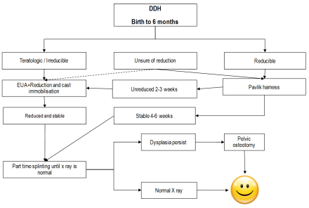
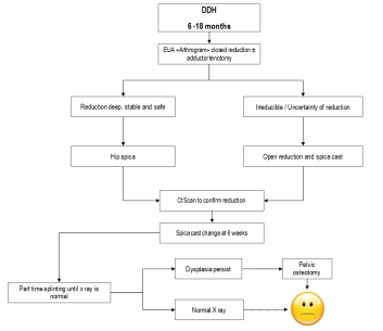
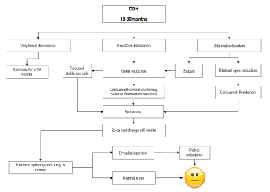
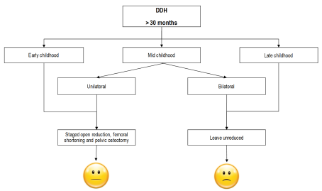
The aim objectives of treatments are to reduce the hip, maintain the reduction while minimising complications. Achieving this depends on the child age, reducibility of the dislocation and available equipments. In general:

 < 6 months

 Pavlik Harness

 6-18 months

 Closed reductions + tenotomy+ hip spica

 18-30 months

 Open reduction ± Pelvic ±femur+ hip spica

 > 30 months

 As above or leave it dislocated.

2.1.6.png

DDH flow chart management (Birth to 6 months). The figure is courtesy of Postgraduate Paediatric Orthopaedic book, Cambridge University Press©
2.1.7.png

DDH flow chart management (6 – 18 months). The figure is courtesy of Postgraduate Paediatric Orthopaedic book, Cambridge University Press©
2.1.8.png

DDH flow chart management (18- 30  months). The figure is courtesy of Postgraduate Paediatric Orthopaedic book, Cambridge University Press©
2.1.9.png

DDH flow chart management (over 30 months). The figure is courtesy of Postgraduate Paediatric Orthopaedic book, Cambridge University Press©

The upper age at which a successful reduction can be carried out has been debated. It is accepted that reduction is recommended for unilateral dislocations in children up to 10 years old and for bilateral dislocations in children up to 8 years. The natural outcome of untreated bilateral dislocations is likely to be better than the results of the reduction of both hips.

Several splints and braces are used to treat children less than 6 months with dislocated hips; the commonest by far is Pavlik harness.  It is relatively easily to apply and most babies tolerate well. It allows motion within the range of stability. This motion is essential for stimulating the growth of acetabulum. The usual position to reduce the hip is 100 degrees of flexion and about 60 degrees of abduction (provided the hip is reduced in this position). Excessive flexion may cause femoral nerve palsy or inferior dislocation while less flexion may cause the hip to re-dislocate. The front leg strap control the amount of flexion while the posterior leg strap controls the abduction. Excessive abduction may cause AVN and less abduction may cause re-dislocation.  It is essential to frequently check the child with PH for hip reduction, hip development, and complications. In dislocated hip, it should be applied full time, however, for dysplasia without hip dislocation, it can used on part time basis. Recognised complications include failure of reduction, AVN  (2.4%), brachial plexus injury, knee dislocation and femoral nerve palsy (2.5%).
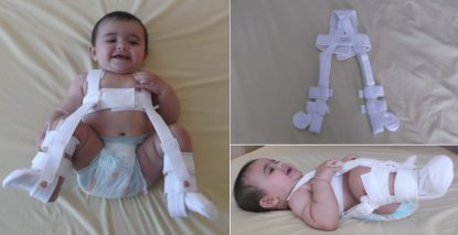
2.1.10.png

A child in a Pavlik Harness.

Video link (child with femoral nerve palsy following Pavlik harness treatment)

https://www.youtube.com/watch?v=v3xEgfa5SH8

Older children or those who failed Pavlik are treated with closed reduction. This is usually performed under general anaesthesia combined with arthrogram to assess the quality of reduction. The range of motion (often called the safe zone) in which the hip remains reduced and the points of dislocation are noted. Adductor tenotomy and / or psoas tenotomy may become necessary to improve the safe zone. The reduced hip is usually maintained in a hip spica (hip abduction of about 45º, flexion of more than 90º and neutral hip rotation).
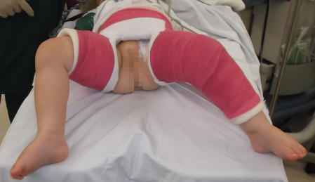
2.1.11.png

A child in a hip spica after open reduction of a dislocated hip. Note the position of the left leg ( 30 flexion ,30 abduction and 30 IR position).

Open reduction is indicated when the reduction is not possible or when the reduction cannot be maintained. Open reduction may be performed from medial approaches (< 1 year) or from an anterior approach (older children). The disadvantages of the medial approach are a limited view of the hip, inability to perform a capsulorrhaphy and the risk of AVN. 

Pelvic osteotomy may be required to improve stability of the reduction. The three commonly used are Salter’s Innominate osteotomy, Pemberton pelvic osteotomy or acetabuloplasty. These are explained in more detailed in a separate chapter. 

Femoral osteotomy may be required especially in older children. This could include shortening, varus, derotation or a combination depending on the stability requirement.
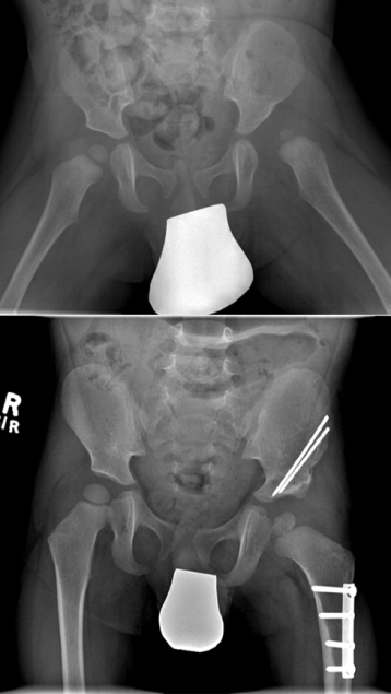
2.1.12.png

Plain radiograph of a child who underwent open reduction, femoral and pelvic (Salter) osteotomy.

It is important to follow up these children until hips become normal as persistent dysplasia is not uncommon and can cause premature osteoarthritis. Surgery for persistent dysplasia is generally indicated if:

  1. The AI fails to improve over 18 months or normalise (AI< 20°) by the age of 4 years
  2. Inadequate acetabular cover (CEA <15°, Uncovering >30%)
  3. Progressive subluxation and instability
  4. Pain with Dysplasia

Based on age, severity of the dysplasia, congruity, and available expertise, the most appropriate procedure is selected. 

Summary for treating residual dysplasia:

 Age

2-6

6- SM

SM

 CR

 Yes

 Yes

 Yes

 No

 Yes

 No

 Size

 N

 L

 N

 S

 N

 S

 Osteotomy


 SO

 PO

 (DO)

 SO

 (TO)

 SA

 (CO)

 GO

 (TO)

 CO

  • SM= Skeletal maturity
  • CR= Concentric reduction
  • S= Small
  • L= Large
  • N= Normal
  • SO= Salter’s Osteotomy
  • PO= Pemberton Osteotomy
  • DO= Dega Osteotomy
  • SA=Shelf Acetabuloplasty
  • CO=Chiari Osteotomy
  • GO=Ganz Osteootmy
2.1.13.png
 
Plain radiograph of a child who underwent open reduction, femoral and acetabuloplasty.

Pelvic osteotomies:

Pelvic osteotomies can be categorised as follows:

1. Reconstructive osteotomies

Re-directional osteotomies
-Single innominate osteotomy (e.g. Salter)
  • Double innominate osteotomy (e.g. Sutherland & Hopf)
  • Triple innominate osteotomy (e.g. Steel, Carlioz, and Tönnis)
  • Bernese periacetabular osteotomy PAO (Ganz)
2.1.14.png
 
Diagram and plain radiograph explaining Salter’s osteotomy.
 
-Reshaping osteotomies
  • Pemberton osteotomy
  • Dega osteotomy
2.1.15.png

Diagram and plain radiograph explaining Pemberton osteotomy.
 
2. Salvage osteotomies
-Chiari osteotomy
-Shelf procedure
  • Reconstructive osteotomies are indicated in hips that have normal joint congruency with no cartilage damage and that reduce concentrically on an abduction-internal rotation (AIR) view.
  • The key acetabular defect in DDH is anterolateral deficiency while in neuromuscular conditions; there is global and posterior deficiency.
  • Re-directional osteotomies in general increase the anterolateral coverage and decrease the posterior coverage and they are useful in DDH but not in neuromuscular dysplasia.
  • Pemberton osteotomy in particular changes the shape and volume of the acetabulum and is primarily useful for a shallow, capacious, or wandering type of acetabulum.
  • The Salter osteotomy, on the other hand, is a re-directional procedure in which the innominate bone is transected completely and then rotated and tilted to provide anterior and lateral coverage of the femoral head without changing the true dimensions of the acetabulum.
  • In an older child or adolescent with an open triradiate cartilage, the triple innominate osteotomy is the procedure of choice. This procedure provides better mobility of the acetabulum and a wider range of coverage; however, it also necessitates more extensive dissection and more than 1 skin incision.
  • After the closure of the triradiate cartilage, Ganz osteotomy is the procedure of choice. This procedure gives extensive mobility to the acetabulum and affords the surgeon the opportunity to translate the hip joint center medially and thereby improve the biomechanical situation. Moreover, PAO can be executed efficiently through a single skin incision, preserves the integrity of the posterior column and so earlier mobilisation can be advised and lastly it doesn’t change the shape of the true pelvis which is a concern in females in the child bearing age.
  • Dega osteotomy increases global coverage including posterior coverage and it is used in neuromuscular hip dysplasia.
  • Salvage osteotomies are done in cases where congruent reduction cannot be achieved and the joint is grossly incongruent.
  • Both Chiari and shelf procedures depend on the interposition of the capsule between the femoral head and either a bone graft in case of shelf procedure or the iliac wing it self with medialisation of the acetabular fragment in Chiari osteotomy

2.1.16.png

Diagram showing various types of commonly used pelvic osteotomies.


Previous
Next

References

  • 1. Bialik, V., G. M. Bialik, et al. (1998). "Prevention of overtreatment of neonatal hip dysplasia by the use of ultrasonography." J Pediatr Orthop B 7(1): 39-42.
  • 2. Jari, S., R. W. Paton, et al. (2002). "Unilateral limitation of abduction of the hip. A valuable clinical sign for DDH?" J Bone Joint Surg Br 84(1): 104-7.
  • 3. Jones, D. (1998). "Neonatal detection of developmental dysplasia of the hip (DDH)." J Bone Joint Surg Br 80(6): 943-5.
  • 4. Salter, R. B. and J. P. Dubos (1974). "The first fifteen year's personal experience with innominate osteotomy in the treatment of congenital dislocation and subluxation of the hip." Clin Orthop Relat Res(98): 72-103.