TOPIC DETAILS

  Team Member Role(s) Profile
SattAR Sattar Alshriyda Section Editor
  • The unique physiological development of the lower limb alignment in children causes significant concerns among parents and doctors.
  • It is important to differentiate between physiological (by far the commonest) and pathological genu varum (bowed leg) or genu valgum.
  • Salenius (1) studied thousands of newly born x-rays and came with curve named after him; Salenius curve.
  • He found that newborn babies have average knee varus of 10° to 15°. As they start walking, the tibio femoral angle progressively moves towards valgus reaching 10° at the average age of 3.5 years. This angle then gradually decreases and settles at 5° of valgus around the age of 8 years
  • Genu varum is likely to be abnormal if:
  1. Presents over the age of 2
  2. Unilateral
  3. Asymmetry of more than 5°
  4. Short stature
  5. Severe varus
  6. > 2 SD of the mean (Salenius Curve)
  7. Obese child
  • Genu varum is likely to be abnormal if:
  1. Unilateral
  2. Severe valgus ( Intermalleolar distance >15 cm at 5 years or 10 cm at 10 years)

3.2.1.png

Salenius curve showing mean tibio femoral angle according to age. Notice the large standard deviation of 8°.  95% of population fall within 2 standard deviation. So single tibio-femoral angle measurement should be interpreted with caution.

3.2.2.png

It is important to have the knowledge and skills to differentiate between pathological and physiological knee mal-alignment.

  1. Blount's disease
  2. Skeletal dysplasia
  3. Tumours
    • Osteochondroma
  4. Metabolic bone diseases
    • Osteogenesis imperfecta
    • Rickets
    • Fluoride poisioning
  5. Focal fibrocartilaginous dysplasia
  6. Infection
  7. Trauma(including iatrogenic)
  1. Renal osteodystrophy
  2. Skeletal dysplasia
    • Morquio syndrome
    • Spondyloepiphyseal dysplasia
  3. Infection
  4. Trauma
    • Proximal tibial metaphyseal fracture (Cozen’s fracture)
  5. Tumours
    • Fibrous dysplasia
    • Osteochondromas
  6. Neuromuscular disease
    • Polio

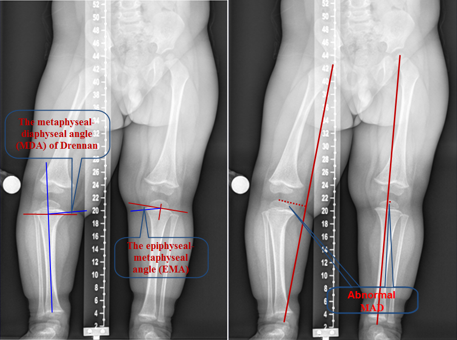
Tibio-Femoral Angle (TFA)

  •  Angle formed by intersection of the two mid-diaphyseal lines of femur and tibia

Mechanical and Anatomical Axis Difference (MAD)

  • Anatomical axis cross at the centre of knee
  • Mechanical axis normally pass 8±7 mm medial to the centre of knee

Metaphyseal-Diaphyseal Angle (MDA)

  • Angle formed by a line connecting the most distal point on the medial and lateral beaks of the proximal tibial metaphysis and a line perpendicular to the anatomic axis (or lateral cortex) of the tibia
  • More than 10°

Epiphyseal-Metaphyseal angle (EMA)

  • Angle formed by a line through the proximal tibial physis parallel to the base of the epiphyseal ossification centre with the most distal point on the medial beak of the proximal tibial metaphysis
  • More than 20°

3.2.3.png

Common radiological measurements when assessing genu varum or valgum; TFA, MAD, MDA and EDA (see text for description).

  • Idiopathic Tibia Vara
  • Abnormal ossification of medial aspect of proximal tibial physis, epiphysis and metaphysis
  • Causes
  1. Actual cause is unknown
  2. Probably caused by a combination of excessive forces on proximal medial metaphysis of tibia and altered enchondral bone formation
  • Radiological assessment
  1. Sharp varus angulation at proximal tibial metaphysis
  2. Widened and irregular physeal line medially
  3. Medially sloped and irregularly ossified epiphysis
  4. Prominent beaking of medial metaphysis with lucent cartilage islands within the beak
  5. Radiological measurements:
    • TFA (varus)
    • MDA > 10°
    • EMA > 20°
  • 2 types
  1. Infantile
  2. Adolescent

3.2.4.png

Blount disease. Varus knees with MDA of 27° and EMA 38°).

  • Under the age of 4
  • Early walker (9 to 10 months old)
  • Bilateral (80%)
  • More prevalent in:
  1. Female
  2. Marked obesity
  3. African origin
  • Associated with:
  1. Lateral thrust of knee
  2. Internal tibial torsion
  3. Leg length discrepancy
  • Langenskiold classification (Fig. 3)
  1. Radiological classification
  2. 6 stages
 3.2.5.png

Langenskiold Classification

Stage 1: Medial beaking, irregular medial ossification with protrusion of the metaphysis. Stage 2: Cartilage fills depression. Progressive depression of medial epiphysis with the epiphysis slopes medially as disease progress. Stage 3: Ossification of the inferomedial corner of the epiphysis. Stage 4: Epiphyseal ossification filling the metaphyseal depression. Stage 5: Double epiphyseal plate (cleft separating two epiphysis). Stage 6: Medial physeal closure.

Bracing

1. Under the age of 3
2. Stage I and II
3. 50% success rate
4. Risk factors for failure:
  • Bilateral
  • Instability
  • Obesity
  • Delayed bracing
Hemiepiphysiodesis
  • Failed bracing treatment
  • Over the age of 3
  • Stage III and above if under the age of 3
Proximal tibial osteotomy is the main stay treatment as all the above are unpredictable.
  • Not as common as infantile Blount’s
  • Above the age of 10
  • Unilateral (80%)
  • May have distal femur deformity
  • More prevalent in:
  1. Overweight
  2. African origin
  • Usually surgical as orthosis is ineffective.
  • Aim to correct the mechanical axis to normal
  • Avoid overcorrection
  • Hemiepiphysiodesis (50 – 70% success rate)
  • Proximal tibial osteotomy
  • Distal femoral osteotomy if femur involved (30%)
  • Stabilisation can be achieved with k-wires, plates or a frame depending on the age of the child, preference of the parents and available resources and expertise.

3.2.6.png

Stabilisation can be achieved using various methods including k-wires, plates or a frame depending on the age of the child, preference of the parents and available resources and expertise.

Previous
Next

References

  • 1. Salenius P, Vankka E. The development of the tibiofemoral angle in children. J Bone Joint Surg Am. 1975 Mar;57(2):259-61.