TOPIC DETAILS

  Team Member Role(s) Profile
SattAR Sattar Alshriyda Section Editor
Akinwande Adedapo Akinwanda Adedapo Segment Author
Mansoor Masroor A Naveed Segment Author
  • MSK infection encompass a wide spectrum of conditions that vary with age, place and host co-morbidity.
  • Septic arthritis, acute and chronic osteomyelitis will be discussed in this section.
  • Septic arthritis refers to “bacterial” invasion of the joint space and the subsequent inflammatory response.
  • Osteomyelitis is an infection of the bone and bone marrow.
  • Delayed or an inappropriate treatment is associated with significant comorbidity.
  • Both conditions are usually caused by similar pathogens (there may be regional variation- always check with your local infectious disease services).

Common causative organisms

 Group

 Causative organism

 Neonate

 Staphylococcus aureus, Group B Streptococcus,

 E. Coli, Haemophilius influenza (now rare due to immunization)

 Early childhood (to 3 years)

 S. aureus, Kingella kingae, Strep pneumonia,

 N. meningitides

 Childhood (3-12 years)

 S. aureus*, Group A β-haemolytic streptococcus

 Adolescent (12-18 years)

 S. aureus*, Group A β-haemolytic streptococcus, Neisseria gonorrhea

 Other risk factor:

  -Sickle cell
  -Foot puncture

 Salmonella species, S. aureus, S. pneumonia, Pseudomonas aeruginosa, S. aureus

Pathogenesis

  • There are several routes where bacteria invade joints
  1. Haematogenous spread
  2. Direct inoculation (surgery or trauma)
  3. From adjacent Osteomyelitis
  • Bacteria release endotoxins and body respond by releasing cytokines, such as tumor necrosis factor and interleukin-1.
  • These cytokines stimulate the release of proteolytic enzymes.
  • This combination of factors leads to destruction of the synovium and articular cartilage.
Clinical Features
  • Constitutional system (fever, malaise, anorexia, irritability and not feeding)
  • Loss of function (Limp, inability to weight bear, pseudo-paralysis in the neonate.
  • Joint effusion, overlying erythema, restricted range of motion.
Investigation
Blood test
  • WCC
  • ESR (doesn't start elevation until 48 hours)
  • CRP may be a better marker in older children as it is elevated ithin 6 hours and peaks over the next 48 hours.
Imaging studies
    • Ultrasound
  1. Non invasive and inexpensive
  2. Operator dependent
  3. Useful tool to detect joint effusion
  4. It can also assess the extra articular structures (joint capsule, periosteum, metaphyseal bone and adjacent soft tissues).
  5. Cannot differentiate between infection and non- infectious inflammatory arthropathy.
1.5.1.png
Figure 1.5.1
Ultrasound images of the right hip in a 3-year-old female. A large effusion is evident (white arrow) with layering suggestive of debris (curved red arrow). Capsular thickening (red arrow) is also evident. Image is courtesy of Postgraduate Paediatric Orthopaedic, Cambridge University Press.
    • MRI
Joint Aspiration
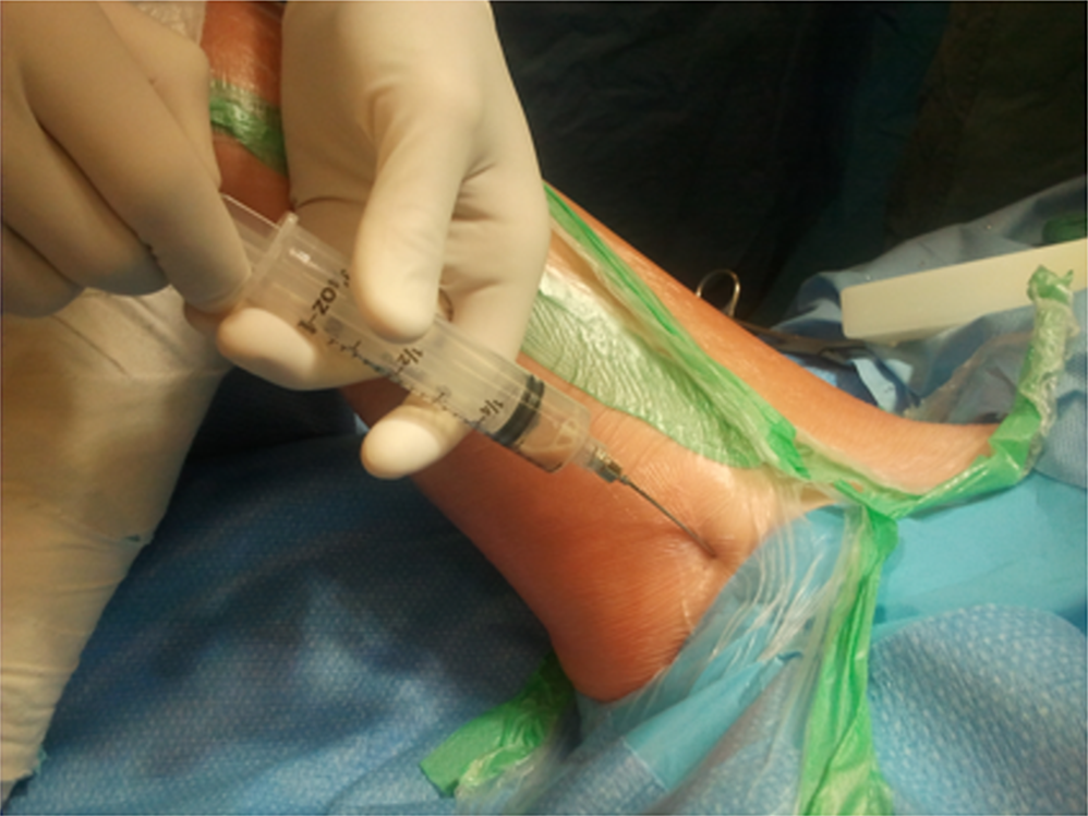
    1. Should be done under complete aseptic technique (first do no harm).
    2. Invasive
    3. Findings can be inconclusive
    4. Defining infection (may vary among various institutes)
  • Positive culture, although can be negative in up to 40% cases of septic arthritis
  • Synovial leukocyte count more than 50,000/mm3 and 75% polymorph presence indicates infection. Or more than 5 pus cell (WCC) per high power field
Differential Diagnosis
  1. Haemarthrosis
  2. Traumatic effusion
  3. Transient synovitis (see below)
  4. Reactive arthritis
  5. Lyme arthritis
  6. Juvenile rheumatoid arthritis ( risk of blindness, if you suspect refer to rheumatologist asap)
  7. Arthritis of acute rheumatic fever
  8. Osteomyelitis
  9. Tumours
  10. Perthe’s disease

Differentiate between Septic arthritis and Transient synovitis:

Transient synovitis is by far more common than septic arthritis; however the latter needs urgent attention.  Kocher et al (1-3) in several studies proposed criteria (named Kocher’s criteria) to help differentiate between them.

He identified four independent predictors to differentiate between Transient synovitis and septic arthritis; Fever > 38.5°c, inability to weight bear, ESR >40, WBC >12,000/mm3.

Kocher’s Criteria

 No. of positive   predictors

 (% risk of septic arthritis)

 Kocher 1999

 (% risk of septic arthritis)

 Kocher 2004

 0

 0.2

  2

 1

 3

  9.5

 2

 40

  35

 3

 93.1

  72.8

 4

 99.6

  93

1.5.2.png

Figure 1.5.2. Medical approach to aspirate a hip Image is courtesy of Postgraduate Paediatric Orthopaedic, Cambridge University Press

Management

Principles of management of acute septic arthritis are:
    1. Adequate and prompt drainage of the joint
    2. Antibiotics to diminish systemic effects of sepsis
    3. Rest joint
  • Antibiotics should be administered once the diagnosis is made. Sample should be sent for gram stain, culture and cell count.
  • The choice of antibiotic therapy depends the local guidelines and adjusted according to the culture results.
  • Ideally this should be in close liaison with microbiologist or an infection control specialist with an interest in MSK.
  • Duration and route of administration is controversial and most units have their own local guidelines.
  • The current trend is IV antibiotic until clinical and biochemical resolution then switch to oral antibiotics for 4-6 weeks of treatment.
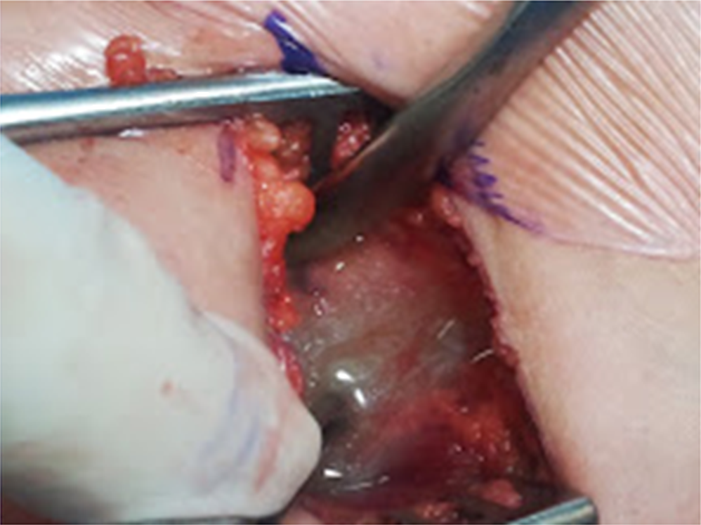
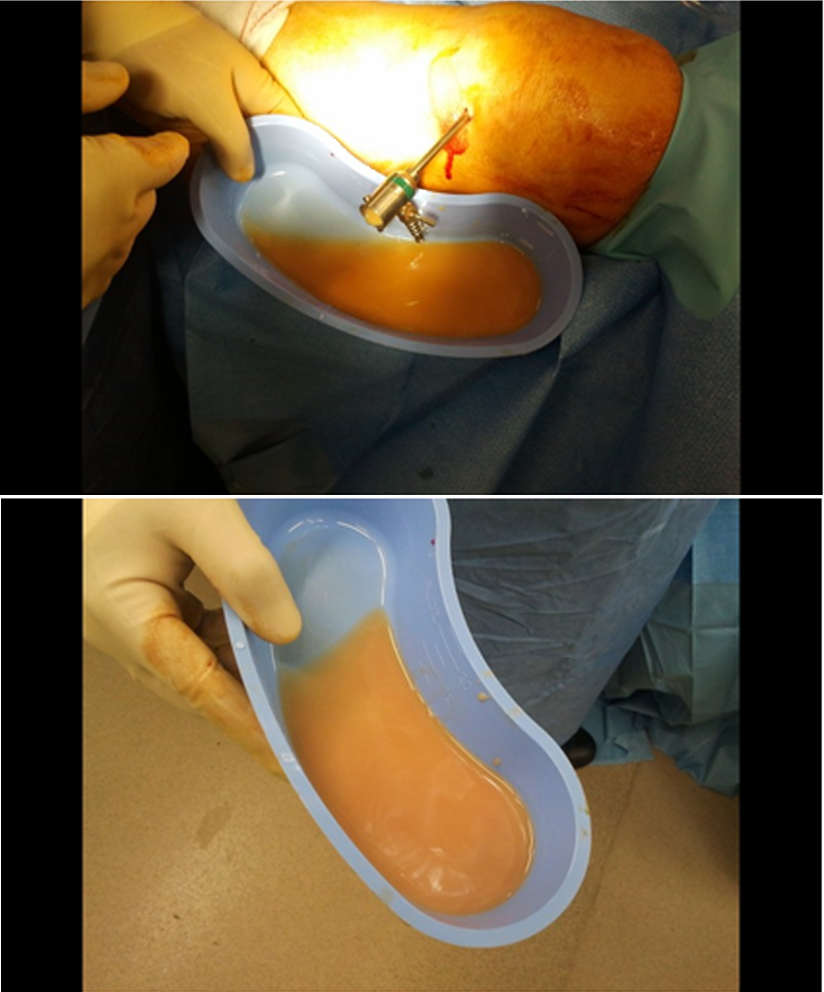
  • Controversy exists about open surgical drainage, arthroscopic drainage and multiple aspirations as good results with all three methods have been reported. The essence of success is an adequate drainage.
  • 1.5.3.png

Figure 1.5.3. Hip joint is full of pus
1.5.4.png

Figure 1.5.4 Arthroscopic washout of an infected knee

Background

  • Osteomyelitis is inflammation of the bone caused by an infecting organism.
  • Despite modern advances, management of osteomyelitis is challenging.
  • Successful treatment requires early diagnosis and a multidisciplinary approach using appropriate antibiotics and aggressive surgical management.
  • Infection may be contained to only one portion of bone or may involve numerous regions (periosteum, cortex, marrow, surrounding soft tissue). Infection can be due to a single organism or polymicrobial.
  • Skeletal distribution: Most commonly involves the lower extremity and order of frequency is Femur 27%, Tibia 26%, Pelvis 9%, Humerus 8%

Classification

  • Osteomyelitis can be classified based on duration of symptoms, mechanism of infection or type of host response:
  • Duration of symptoms:
  1. Acute
  2. Subacute
  3.  Chronic
  • Mechanism:
  1. Exogenous (open fracture, surgery, local spread)
  2. Haematogenous (bacteraemia)
  • Host response:
  1. Pyogenic
  2. Non pyogenic
  • Chronic osteomyelitis is generally classified by Cierny & Mader based on host’s health status (A. normal, B. compromised, C. prohibitive) and bone site involvement (I. medullary, II. cortical, III. localised and IV. diffuse) into possible twelve categories.

Acute haematogenous Osteomyelitis (AHOM):

  • It is the commonest form in children; probably because:
  1. Metaphyseal ends of rapidly growing long bones have sluggish turbulent blood flow due to numerous vascular loops.
  2. Relative paucity of reticuloendothelial cells adjacent to the physis
  3. Fewer phagocytosis cells in the metaphysis (than epiphysis or diaphysis)
  4. Lack of basement membrane in synovial capillaries
  • Age distribution for AHOM in children is bimodal
  • Affecting children less than 2 and 8-12 years.
  • In the former, the metaphysis is usually intra-intra-articular and may present as septic arthritis.
  • In the latter, the physis prevents spread of infection to the epiphysis and infection usually spreads towards diaphysis.

 1.5.5.png

Figure 1.5.5 Brodie’s abscess treated with drainage and curettage

Pathogenesis

  • Bacterial seeding leads to inflammatory reaction and thrombosis of endosteal blood vessels, leading to local ischaemic necrosis of bone and subsequent abscess formation. It is often called a Brodie’s abscess (figure 1.5.5)
  • As metaphyseal cortex is thinned, pus ruptures to form a subperiosteal abscess, further depriving the cortex of its blood supply.
  • If process continues the segment of cortex becomes necrotic and forms a sequestrum.
  • The periosteum though retains its osteogenic property and tries to make new bone around the sequestrum, which is called an involucrum (sequestrum and involucrum are hallmarks of chronic osteomyelitis) (figure 1.5.6). 
  • Osteomyelitis of the metaphysis may lead to thrombosis of transphyseal vessels leading to severe osteonecrosis of the epiphysis resulting in angular deformity or limb shortening.
  • However once the ossific nucleus appears the epiphyseal and metaphyseal blood supply becomes independent and the physis acts as a barrier to spread.
1.5.6.png

Figure 1.5.6 Chronic osteomyelitis. The entire diaphysis became a sequestrum. The involucrum can be seen engulfing the diaphysis. Image is courtesy of Postgraduate Paediatric Orthopaedic, Cambridge University Press

Causative organisms

  • Staph aureus is the most common pathogen in children followed by group B streptococcus (infection in healthy infants 2-4 week old) and gram-negative coliforms.
  • Sickle cell associated infection tends to be more diaphyseal than metaphyseal.
  • MRSA infections have recently been reported in upto 25% cases in one of the centres.

Diagnosis and Investigations

  • A good history (pre, peri and postnatal) and thorough physical examination is necessary. In infants there may be minimal clinical findings and fever and malaise may not be present in the earlier stages though pain, swelling and tenderness are common findings.
  • Soft tissue swelling may start 2-3 days after infection has developed.

Blood tests:

  • FBC is often normal but ESR and CRP levels are usually elevated.
  • Blood culture ( may need to be repeated if spiking temperature continues)- positive in 50%

Imaging studies:

  • Radiographs may only show soft tissue reaction at 1-3 days but skeletal changes like subperiosteal reaction and bony destruction becomes visible at 10-12 days into infection.
  • Tc99 scan can confirm diagnosis in 90-95% patients within 24-48 hours of onset and a negative Tc99 scan effectively rules out presence of acute osteomyelitis.

Bone biopsy/ Local swabs

  • Bone biopsy must be performed through uninvolved tissue.
  • Cultures of the sinus tract may be useful if S aureus and Salmonella species are isolated (4, 5)

Treatment

Acute Haematogenous Osteomyelitis

  • Most hospitals have a locally agreed protocol which is based on commonly isolated organisms.
  • In our centre, for acute uncomplicated osteomyelitis a two weeks course of intravenous antibiotic is followed by 4 weeks of oral antibiotic.
  • Empirical treatment with Cefatriaxone (50mg/kg) and Flucloxacillin (50mg/kg) is started after all cultures are taken.
  • It is valuable to identify causative organisms as early as possible and adjust antibiotics accordingly.
  • The timing for conversion to oral antibiotics is also dependent upon the resolution of clinical signs (temperature, overlying erythema and tenderness), CRP and WCC returning to baseline and availability of intravenous access.
  • Various treatment protocols (shorter and longer) have been published with similar success rates (6-8).

Surgery

  • Large abscesses (>2cm) should be considered for drainage (8, 9).
  • Surgical treatments for chronic osteomyelitis are better guided by Cierny types (Table 1.5.3)(10)
  • Type 1 and 2 do not usually require surgical treatment, whereas stage 3 and 4 do.
  • Surgical treatments encompass drainage, debridement of dead tissues, management of dead space, adequate soft-tissue coverage, restoration of blood supply and protection against fracture (cast and external fixators are valuable).
  • Type C hosts, treatment may be more harmful than the osteomyelitis itself and better to control rather than trying to cure it

Cierny Staging

  Anatomical type:


  I

 Medullary

 Endosteal surface involved

  II

 Cortical Superficial

 Coverage defect

  III

 Localised

 Localized cortical sequestrum

  IV

 Diffuse

 I+II+III+ mechanical instability

  Host type


  A

 Normal

 Immunocompetent/ good local vascularity

  B

 Compromised

 Local/systemic factors compromise immunity

  C

 Prohibitive

 Morbidity with treatment/ poor prognosis


Previous
Next

References

  • 1. Kocher MS, Mandiga R, Murphy JM, Goldmann D, Harper M, Sundel R, et al. A clinical practice guideline for treatment of septic arthritis in children: efficacy in improving process of care and effect on outcome of septic arthritis of the hip. J Bone Joint Surg Am. 2003 Jun;85-A(6):994-9.
  • 2. Kocher MS, Mandiga R, Zurakowski D, Barnewolt C, Kasser JR. Validation of a clinical prediction rule for the differentiation between septic arthritis and transient synovitis of the hip in children. J Bone Joint Surg Am. 2004 Aug;86-A(8):1629-35.
  • 3. Kocher MS, Zurakowski D, Kasser JR. Differentiating between septic arthritis and transient synovitis of the hip in children: an evidence-based clinical prediction algorithm. J Bone Joint Surg Am. 1999 Dec;81(12):1662-70.
  • 4. Howard CB, Einhorn M, Dagan R, Yagupski P, Porat S. Fine-needle bone biopsy to diagnose osteomyelitis. J Bone Joint Surg Br. 1994 Mar;76(2):311-4.
  • 5. Perry CR, Pearson RL, Miller GA. Accuracy of cultures of material from swabbing of the superficial aspect of the wound and needle biopsy in the preoperative assessment of osteomyelitis. J Bone Joint Surg Am. 1991 Jun;73(5):745-9.
  • 6. Jagodzinski NA, Kanwar R, Graham K, Bache CE. Prospective evaluation of a shortened regimen of treatment for acute osteomyelitis and septic arthritis in children. Journal of pediatric orthopedics. [Controlled Clinical Trial]. 2009 Jul-Aug;29(5):518-25.
  • 7. Peltola H, Paakkonen M, Kallio P, Kallio MJ. Short- versus long-term antimicrobial treatment for acute hematogenous osteomyelitis of childhood: prospective, randomized trial on 131 culture-positive cases. The Pediatric infectious disease journal. [Randomized Controlled Trial Research Support, Non-U.S. Gov't]. 2010 Dec;29(12):1123-8.
  • 8. Dartnell J, Ramachandran M, Katchburian M. Haematogenous acute and subacute paediatric osteomyelitis: a systematic review of the literature. The Journal of bone and joint surgery British volume. [Review]. 2012 May;94(5):584-95.
  • 9. Connolly SA, Connolly LP, Drubach LA, Zurakowski D, Jaramillo D. MRI for detection of abscess in acute osteomyelitis of the pelvis in children. AJR American journal of roentgenology. 2007 Oct;189(4):867-72.
  • 10. Cierny G, 3rd, Mader JT, Penninck JJ. A clinical staging system for adult osteomyelitis. Clin Orthop Relat Res. 2003 Sep(414):7-24.