TOPIC DETAILS

  Team Member Role(s) Profile
SattAR Sattar Alshriyda Section Editor
  • The severity of cerebral palsy (CP) can vary from subclinical findings to very severe and disabling deformities.
  • Symptoms and signs usually get worse as the child grows. Moreover, children develop compensatory or secondary deformity to improve their functional capability. This makes surgical decisions somehow unpredictable.
  • The introduction of Botox treatment and gait analysis has changed the approach to treating children with CP from addressing single problems one at a time (commonly called the Birthday Syndrome) to a single event multilevel surgical approach (SEMLS) where multiple deformities are addressed simultaneously.
  • There are obvious benefits of SEMLS in carefully selected patients (one anaesthetic, one admission, better outcomes, less cost) however, it can be less predictable and the risk of surgery is higher.
  • Even in SEMLS, surgeons occasionally do not correct every deformity particularly if they are minor ones.  Future “tidying operation” may become necessary to deal with uncorrected deformities if they impair function.
1.8.2.1.png
 
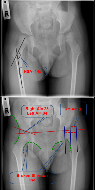
Plain pelvic x-ray of a child with CP showing commonly measured parameters. NSA is the angle between the femoral shaft and neck (125-135°). Reimer’s migration index (RMI) is the percentage of uncovered head (= width of the uncovered head / the width of the head X 100). Normal RMI is less than 30%. Traditionally, RMI between 30-50% is considered as an early subluxation, > 50% as a moderate subluxation and > 90% as a dislocation.

Orthopaedic procedures that are useful in CP can be divided into:

Soft tissue procedures:

  1. Tenotomy
  2. Tendon transfer

Both inevitably weaken the tendons on a permanent basis, therefore bony procedures are often preferred where possible.

Bony Surgery: 

Bony surgery is required to realign the bones and restore optimum lever functions. The following bony surgery is commonly performed:

  • Long bone osteotomy:
  1. Shortening (e.g. proximal femur)
  2. Lengthening (Calcaneum)
  3. Derotation (femur or tibia)
  4. Varus (femur)
  5. Extension (proximal or distal femur)
  6. Combined (femoral VDRO).
  • Pelvic  osteotomy (e.g. Dega osteotomy)
  • Epiphysiodesis
  • Arthrodesis
  • Salvage surgery such as excision of the proximal femur
  • Hip abnormalities are common (> 80 % in GMFCS level V)
  • Adduction contractures (<30°), excessive femoral anteversion, coxa valga and acetabular dysplasia can lead to hip dislocation.
  • Hip abduction of <30° is a herald of joint subluxation.
  • Radiographically, 4 parameters are used to evaluate the hip in children with CP;(1) the femoral neck-shaft angle (NSA),(2) Shenton’s line, (3)Reimer’s migration index (RMI) and (4)the acetabular index (AI)

Surgical treatment

This may involve any or all of the followings depending on the severity:

  1. Adductor(s) release (commonly involves adductor longus, Gracilis and adductor brevis). It is usually indicated if abduction < 30°  
  2. Flexion contracture is usually indicated when FFD > 30°. This can be achieved by intramuscular lengthening of the iliopsoas tendon over the pelvic brim through an anterior hip (Smith-Peterson) approach in ambulant child. This preserves muscle power and prevents excessive weakness associated with dividing the tendon from a medial approach. If femoral osteotomy is planned at the same time, adding extension to the osteotomy may be adequate so as not to need dividing the iliopsoas.
  3. Femoral osteotomy to correct excessive femoral anteversion (de-rotation), coxa valga (varus); flexion contracture (extension) or collectively to correct all these deformity as they often co-exist
  4. Pelvic osteotomy through an anterior hip approach. In contrast to DDH, the acetabular deficiency in a neurogenic hip is usually posterior and lateral or global rendering Slater’s osteotomy not useful (may be harmful). Dega osteotomy (or one of its modification) is usually indicated when there is significant dysplasia (AI> 20°)

1.8.2.3.png

Surgical steps in Dega osteotomy. 1 & 2 osteotomy around the superior posterior acetabular rim; open the space using laminar spreader (4). Bone graft harvested from the iliac crest (3&5) and divided into wedges (6) which are impacted in the osteotomy (7).

1.8.2.4.png

A child with neurogenic hip dislocation who underwent hip recon struction. 

In children with CP, the knee can be involved in the following pathological process:

1. Hamstring contracture:

  • This is usually manifested by short step, hyperlordosis and can be assessed by measuring the popliteal angle.
  • It is common in patients with diplegia and quadriplegia.
  • Medial hamstrings (semitendonosus, semimembranosus and gracilis) are usually lengthened.
  • In severe deformity, a recession of the biceps femoris (lateral hamstring) may also be indicated.
  •  Anterior hemi-epiphysiodesis (in younger patients, < 30° of FFD) or (extension osteotomy of the distal femur) may be added to provide more correction or to avoid weakening the hamstring.
  • Complications of the above procedures include damage to the sciatic or common peroneal nerve, genu recurvatum and increased anterior pelvic tilt.

2. Tight rectus femoris:

  • It results in anterior knee pain, stiff knee gait and patella alta.
  • It impairs knee clearance during swing phase.
  • The rectus tendon can be recessed or transferred to one of the medial hamstring. There is no evidence to suggest transfer is superior to recession only.
  • Patalla alta is usually treated by patellar pull down procedure or Y-V patellar tendon shortening

1.8.2.5.png

Extension osteotomy of distal femur in a child with flexion contracture of the knee

1.8.2.6.png

Anterior hemi-epiphysiodesis of the distal tibia to correct fixed flexion deformity of the knee

Four foot and ankle deformities are commonly associated with CP:

  1. Equinus
  2. Equinovarus
  3. Equinovalgus
  4. Hallux valgus

Equinus

  • True equinus is usually caused by gastrocnemius with or without soleus
  • Silfverskiöld test is used to differentiate between the two.
  • The degree of dorsiflexion is measured with the knee flexed and extended. Flexing the knee relaxes the gastrocnemius and test the tightness in soleus. Extending the knee test tightness in both muscles.
  • Lengthening gastrocnemius preserves the power of push off (by the soleus) and is less likely to result in overcorrection ( which is more problematic); however, there is a higher recurrence rate.

Equinovarus

  • This is most often seen in the hemiplegic palsy.
  • Tightness in tibialis posterior and or anterior as well as tendoachilles is implicated.
  • The confusion test is proposed to help to determine which muscle is the main culprit.
  • With active hip flexion, if the forefoot supinated, the tibialis anterior is likely involved while pure dorsiflexion suggests involvement of the tibialis posterior. However, this has been contested based on gait analysis studies.
  • When the deformity is not rigid, muscle balance around the foot and ankle can resolve the problem
  • Split tendon transfer of either tibialis posterior or anterior may be required to balance the forces and prevent recurrence. Our preferred method is split tibialis anterior transfer to the cuboid, tibialis posterior and tendoachilles lengthening then rigid AFO.
  • In severe and rigid deformity, lateral calcaneum shift osteotomy, first ray elevation may be necessary

Equinovalgus

  • This is more common in diplegics and quadriplegics and it is usually caused by tight tendoachilles, spastic peroni and weak tibialis posterior or various mix of them.
  • In ambulant children, it is preferable to control by AFO, however, in severe deformity AFO may not fit very well and cannot be tolaerated.
  • Surgical treatment involves lengthening the contracted muscles (tendoachillis and peroneus brevis) and lateral column lengthening through the calcaneum. There are other alternatives depending on the severity of the deformity and the state of other joints.
1.8.2.7.png

Clinical photographs of a CP child with severe equinus valgus. 

1.8.2.8.png

These are the preoperative and postoperative x-rays of the boy shown in figure 1.8.1.3 who presented with severe equinus valgus. He was treated with tendoachilles lengthening, peroni lengthening and lateral column lengthening.

Hallux valgus

  • It is usually associated with equinovalgus foot and external tibial torsion
  • Arthrodesis is the most reliable as other procedures have a high recurrence rate.

 1.8.2.9.png

This CP child presented with severe left hallux valgus

Previous
Next