TOPIC DETAILS

  Team Member Role(s) Profile
SattAR Sattar Alshriyda Section Editor
  • Bipartite patella is the result of failure of fusion of a secondary ossification centre.
  • In 70% of the cases, the accessory patellar portion is on the superolateral pole.

3.6.1.1.png

Saupe’s classification for accessory patella. Picture is courtesy of Postgraduate Paediatric Orthopaedics.

  • Incidental finding in approximately 2% to 3% of normal people.
  • It can cause activity related pain around the bipartite fragment.
  • Direct palpation of the fragment can reproduce the pain.
  • X-rays and an MRI scan can assist in the diagnosis.

3.6.1.2.png

X-ray showing a bipartite patella.

  • Treatment for a symptomatic bipartite patella is initially conservative.
  • It includes activity modification, rest, NSAID and use of knee immobilizer.
  • In case of failure of conservative treatment surgical lateral release with or without fragment excision can provide pain relief (1).
  • It is a rare condition manifested at birth with permanent and irreducible dislocation of the patella.
  • It is combined with flexion contracture of the knee, genu valgum and external tibial torsion.
  • It is believed to be the result of failure of the quadriceps-containing myotome to undergo internal rotation, that normally occurs within the first prenatal trimester (2).

3.6.2.1.png

Clinical photograph and plain radiographs showing patellar dislocation. Noted it is dislocated in flexion indicating short quadriceps tendon.

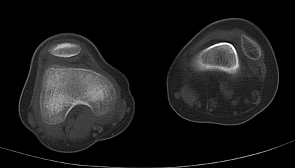
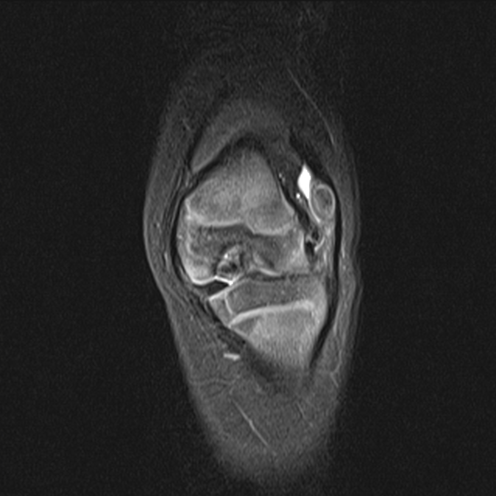
  • There is fixed lateral dislocation of a hypoplastic patella, lack of active knee extension and absence of the patella from intertrochlear fossa.
  • Congenital patellar dislocation can be associated with Larsen’s syndrome, arthrogryposis, myelomeningocele, Down syndrome, nail-patella syndrome, Beckwith-Wiedemann syndrome, diastrophic dysplasia and PFFD.
  • Ultrasound can be used to locate the patella and confirm the diagnosis.
  • MRI can be used as part of preoperative assessment and planning.
3.6.2.2.png

CT scan showing a hypoplastic patella dislocated laterally.
3.6.2.3.png

MRI scan showing a hypoplastic patella dislocated laterally and external torsion of the tibia in relation to the femur.

  • The treatment is surgical realignment early in life (6-12 months).
  • It includes extensive lateral release and advancement of the VMO distally and medially, IT band transverse division and quadriceps lengthening by either V-Y plasty or femoral shortening (3).
  • Patellofemoral (PF) pain is in adolescents.
  • Girls are more frequently affected than boys.
  • Most patients have symptoms bilaterally, although one side may be less symptomatic than the other.
  • The pain is localized at the anterior aspect of the patella.
  • The causes of PF pain are multiple (4-6):
  1. Biologic (chondromalacia patella)
  2. Mechanical (femoral, tibial torsion, excessive foot pronation, patellar malalignement).
  3. Psychosomatic.

Anterior knee pain that increases with climbing, squatting or prolonged sitting.

  • Physical examination:
    • Behavioral assessment
    • Gait assessment
    • Patient sitting (patella baja/alta or subluxation, J-sign)
    • Patient supine ( effusion, patellar mobility, region of tenderness, Q-angle)
    • Patient prone ( femoral anteversion, tibial torsion)
  • Imaging:
    • X-rays (patella alta/baja, patellar tilt/subluxation, trochlear dysplasia)
    • CT (tibial tuberosity-trochlear groove distance, abnormal >20 mm)
    • MRI (articular cartilage abnormalities, joint congruity)

The symptoms of most of these patients resolve over time (7, 8).

  • Mainly conservative:
    • Physical therapy (core stability, VMO strengthening) (9)
    • Activity modification
    • Emotional stress management
    • Pain management
    • Knee orthoses/taping
    • Weight management
  • Surgical to treat instability with associated pain
  • Lateral retinacular when the lateral retinaculum is tight
  • Proximal re-alignment procedures (medial reefing, VMO advancement)
  • Tibial tuberosity transfer (Fulkerson, Maquet, Elmsie and Trillat) in skeletally mature patients
  • Patellar tendon medicalization (Roux–Goldthwaite procedure) or semitendinosus tenodesis of the patella (Galeazzi procedure) in skeletally immature patients
Previous
Next

References

  • 1. Weckstrom M, Parviainen M, Pihlajamaki HK. Excision of painful bipartite patella: good long-term outcome in young adults. Clin Orthop Relat Res. 2008 Nov;466(11):2848-55.
  • 2. Stanisavljevic S, Zemenick G, Miller D. Congenital, irreducible, permanent lateral dislocation of the patella. Clin Orthop Relat Res. 1976 May(116):190-9.
  • 3. Gordon JE, Schoenecker PL. Surgical treatment of congenital dislocation of the patella. J Pediatr Orthop. 1999 Mar-Apr;19(2):260-4.
  • 4. Fairbank JC, Pynsent PB, van Poortvliet JA, Phillips H. Mechanical factors in the incidence of knee pain in adolescents and young adults. J Bone Joint Surg Br. 1984 Nov;66(5):685-93.
  • 5. Hinton RY, Sharma KM. Acute and recurrent patellar instability in the young athlete. Orthop Clin North Am. 2003 Jul;34(3):385-96.
  • 6. Carlsson AM, Werner S, Mattlar CE, Edman G, Puukka P, Eriksson E. Personality in patients with long-term patellofemoral pain syndrome. Knee Surg Sports Traumatol Arthrosc. 1993;1(3-4):178-83.
  • 7. Sandow MJ, Goodfellow JW. The natural history of anterior knee pain in adolescents. J Bone Joint Surg Br. 1985 Jan;67(1):36-8.
  • 8. Nimon G, Murray D, Sandow M, Goodfellow J. Natural history of anterior knee pain: a 14- to 20-year follow-up of nonoperative management. J Pediatr Orthop. 1998 Jan-Feb;18(1):118-22.
  • 9. Crossley K, Bennell K, Green S, McConnell J. A systematic review of physical interventions for patellofemoral pain syndrome. Clin J Sport Med. 2001 Apr;11(2):103-10.