TOPIC DETAILS

  Team Member Role(s) Profile
SattAR Sattar Alshriyda Section Editor, Segment Author

The aim of treatment is to prevent additional slippage by providing mechanical stability using screws or pins while avoiding the complications of AVN and chondrolysis. The choice of treatment depends on the type of slip, its severity, and surgical expertise. Figure 6 summarises the authors’ recommendations.

The recommend treatment of mild and moderate slip is by pinning-in-situ (PIS). However, there is significant controversy on treating severe slip, pinning in situ can be technically difficult if not impossible. Forceful reduction increases the risk of AVN and should be avoided. The options are either pinning in situ (if feasible) or corrective osteotomy as the primary treatment. The remodeling potential is not always predictable and a re-alignment procedure may be necessary with the former option. AVN is uncommon in stable slips, makes the latter option is not a popular choice in stable slips even when it is severe. In contrast to unstable slips where the risk of AVN is high (47%) and may justify open reduction and stabilisation.

fig 6.png

Flow chart of SUFE treatment. Courtesy of Cambridge University Press.

Several studies have tried to identify the best time to operate on SUFE, all suffer from flaws.

Loder in his classic paper (Loder 1993) noted more AVN in patients treated within 48 hours (7/8 versus 7/21). In contrast Peterson (Peterson, Weiner et al. 1997) showed stabilisation of SUFE within 24 hours led to less AVN (3/42=7%) in comparison with those stabilised after 24 hours (10/49=20%).

Phillip’s (Phillips, Griffiths et al. 2001) reported no single AVN in 16 unstable slips who were treated with 24 hours. In a study of 82 children (117 SUFEs), Kalogrianitis (Kalogrianitis, Tan et al. 2007) showed that AVN developed in 50% (8/16) of the unstable SUFEs. All but one were treated between 24 and 72 hours after symptom onset. They recommended the operation should be done within 24 hours or after 7 days but not in between.  

In a metanalysis of 5 studies, (Lowndes 2009) analysed data from 130 unstable SUFEs ; 56 were treated within 24 hours and 74 were treated after 24 hours of symptoms onset. They found that the odds for developing AVN if treatment occurs within 24 hours were half than those of developing AVN if treatment occurs beyond this point. (OR=0.50; 95% CI: 0.09–2.92; P: 0.441), a statistically non significant findings.

Four operations are in use for primary open reduction and fixation of the slip; Fish osteotomy (Fish 1984), Dunn’s Osteotomy (Dunn and Angel 1978),  Ganz surgical dislocation of the hip (Ganz, Gill et al. 2001) and Parsch technique (Parsch, Weller et al. 2009). Ganz surgical dislocation is also called modified Dunn’s osteotomy. They correct the slip at the subcapital level; however, they differ in the approaches.In Fish osteotomy and Parsch technique, Smith-Peterson anterior approach to the hip is used and it does not involve trochanteric osteotomy. Whereas in Dunn’s and Ganz osteotomy, lateral approach is used and the greater trochanter is osteotomised through the growth plate (Dunn’s osteotomy) or by flip trochanteric osteotomy (Ganz technique). The reported AVN rates are:

  • Fish osteotomy (3/66) (4%).
  • Dunn’s Osteotomy (9/73) (12%)
  • Ganz osteotomy (0/20) (0%).
  • Parsch technique (3/64)

The success of the above approaches is closely related to protecting (may be restoring) the blood supply to the femoral head which comes mainly from the posterior portion of the extra-capsular anastomotic ring. The incidences of AVN reported in the original reports are not comparable as the first two operations probably combined stable and unstable slips. Very few centres have reproduced Ganz results (Sankar, McPartland et al. 2010; Alves, Steele et al. 2013). Since it is publication, the Parish technique has become popular but comparable results from other centres remain to be seen.

Current recommendation for patients who are willing to accept a higher risk of AVN to correct a severe slip or slips which cannot be pinned, is the Parish technique for unstable one ( that does not reduce spontaneously) and Ganz osteotomy for stable ones.

fig 7.png

Severe SUFE in an overweight child treated with Dunn’s osteotomy. Notice the low uptake on the bone scan which is consistent with an AVN.

fig 8.tif.png

fig 8b.png

Severe SUFE with MRI evidence of AVN. There  is  high  STIR  signal within  the  left  femoral  head  consistent with AVN. This patient was treated with surgical dislocation and intra-operative pictures showed bleeding from the femoral head.

The risk of contralateral slip varies from 18 to 60% which could justify prophylactic PIS of the contralateral hip even if it is asymptomatic. However, this is not free of risk and it should be weighed against the benefit.

In a review of 50 children with unilateral SUFE, (Stasikelis, Sullivan et al. 1996) tried to identify predictors for future contralateral slip. They found the modified Oxford bone age strongly correlated with the risk of development of a contralateral slip; contralateral slip developed in 85% of patients with a score of 16, in 11% of patients with a score of 21, and in no patient with a score of 22 or more. The modified Oxford bone age is based on appearance and subsequent fusion of the iliac apophysis, femoral capital physis, greater and lesser trochanter.

The posterior slip angle (PSA) was proposed by  (Phillips, Phadnis et al. 2013) as a reliable predictor for contralateral slip. They examined 132 patients with SUFE and found the PSA was significantly higher in children who subsequently developed contralateral slip ( 17.2° ± 5.6°; N=42 vs. 10.8° ± 4.2°; N=90; P=0.001). If a posterior sloping angle of 14° were used as an indication for prophylactic fixation, 35 (of 42 = 83.3%) would have been prevented, and 19 ( of 90 = 21.1%) would have been pinned unnecessarily.

The followings may aid decision making (Alshryda, Jones et al. 2014):

  1. Age of the child (< 10 years is associated with a higher risk of bilaterality).
  2. Slips associated with renal osteodystrophy and endocrine disorders (a  high incidence of bilaterality)
  3. Poor compliance of the child and family.

The nature of current slip (very bad slip occurred over a very short period of time may justify pinning the other side)
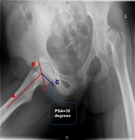
Fig 10.png

The posterior sloping angle (PSA) measured by a line (A) from the center of the femoral shaft through the center of the metaphysis,. A second line (B) is drawn from one edge of the physis to the other, which represents the angle of the physis. Where lines A and B intersect, a line (C) is drawn perpendicular to line A. The PSA is the angle formed by lines B and C posteriorly as illustrated.

  1. AVN
  2. Chondrolysis: Chondrolysis is the rapid and progressive loss of articular cartilage seen in some SUFEs. The cause is unknown; however, few theories have postulated an autoimmune phenomenon or some interference with cartilage nutrition. Risk factors leading to chondrolysis include immobilisation in a cast, unrecognised pin penetration and severe SUFE rapid and progressive loss of articular cartilage after PIS. This may be due to pin penetration or protruded screw, but it has been also reported in other situations. Clinically patient develops increasing pain and stiffness and the x ray shows a decrease in the apparent joint space of more than two millimeters from that of the contralateral hip.
  3. Residual deformity and femoro-acetabular impingement
  4. Osteoarthritis (OA)
  5. Leg length discrepancy (LLD)
Previous
Next

QUESTION 1 OF 1

105.A 15-year-old boy  had a pinning in situ for moderate slipped capital epiphysis 4 years ago(Figure 1). He presents to the clinic with groin pain which is worse on activity. He finds riding a bicycle is particularly painful. The pain gots worse after a recent long haul flight to Australia. Examination showed an externally rotated limb but he is able to correct it. Flexion is limited to 80 degrees before obligatory external rotation happens. Blood tests are normal.
The most appropriate treatment is:

Sattar 3.png

Figure 1.Anteroposterior (AP) pelvis

QUESTION ID: 1297

1. Hip arthroscopy to shave cam lesion
2. Local anaesthetic and steroid injection to confirm the source of pain
3. Removal of metalwork then MRI scan to rule out AVN
4. Southwick or Imhauser femoral osteotomy
5. Surgical dislocation and femoral osteoplasty

References

  • 1. Alshryda, S., S. Jones, et al. (2014). Postgraduate Paediatric Orthopaedics: The Candidate's Guide to the FRCS (Tr and Orth) Examination. Cambridge, Cambridge University Press.
  • 2. Alves, C., M. Steele, et al. (2013). "Open reduction and internal fixation of unstable slipped capital femoral epiphysis by means of surgical dislocation does not decrease the rate of avascular necrosis: a preliminary study." J Child Orthop 6(4): 277-83.
  • 3. Dunn, D. M. and J. C. Angel (1978). "Replacement of the femoral head by open operation in severe adolescent slipping of the upper femoral epiphysis." J Bone Joint Surg Br 60-B(3): 394-403.
  • 4. Fish, J. B. (1984). "Cuneiform osteotomy of the femoral neck in the treatment of slipped capital femoral epiphysis." J Bone Joint Surg Am 66(8): 1153-68.
  • 5. Ganz, R., T. J. Gill, et al. (2001). "Surgical dislocation of the adult hip a technique with full access to the femoral head and acetabulum without the risk of avascular necrosis." J Bone Joint Surg Br 83(8): 1119-24.
  • 6. Kalogrianitis, S., C. K. Tan, et al. (2007). "Does unstable slipped capital femoral epiphysis require urgent stabilization?" J Pediatr Orthop B 16(1): 6-9.
  • 7. Loder, R. T. and M. L. Greenfield (2001). "Clinical characteristics of children with atypical and idiopathic slipped capital femoral epiphysis: description of the age-weight test and implications for further diagnostic investigation." J Pediatr Orthop 21(4): 481-7.
  • 8. Loder, R. T. R., B. S;Shapiro, P. S;Reznick, L. R;Aronson, D. D; (1993). "Acute slipped capital femoral epiphysis: the importance of physeal stability." J Bone Joint Surg Am 75(8): 1134-40.
  • 9. Lowndes, S. K., A.;Emery, D.;Sim, J.;Maffulli, N. (2009). "Management of unstable slipped upper femoral epiphysis: a meta-analysis." Br Med Bull 90: 133-46.
  • 10. Montgomery, R. (2009). "Slipped upper femoral epiphysis." Orthopaedic and Trauma 23:3(June): 169-183.
  • 11. Parsch, K., S. Weller, et al. (2009). "Open reduction and smooth Kirschner wire fixation for unstable slipped capital femoral epiphysis." J Pediatr Orthop 29(1): 1-8.
  • 12. Peterson, M. D., D. S. Weiner, et al. (1997). "Acute slipped capital femoral epiphysis: the value and safety of urgent manipulative reduction." J Pediatr Orthop 17(5): 648-54.
  • 13. Phillips, P. M., J. Phadnis, et al. (2013). "Posterior sloping angle as a predictor of contralateral slip in slipped capital femoral epiphysis." J Bone Joint Surg Am 95(2): 146-50.
  • 14. Phillips, S. A., W. E. Griffiths, et al. (2001). "The timing of reduction and stabilisation of the acute, unstable, slipped upper femoral epiphysis." J Bone Joint Surg Br 83(7): 1046-9.
  • 15. Sankar, W. N., T. G. McPartland, et al. (2010). "The unstable slipped capital femoral epiphysis: risk factors for osteonecrosis." J Pediatr Orthop 30(6): 544-8.
  • 16. Southwick, W. O. (1984). "Slipped capital femoral epiphysis." J Bone Joint Surg Am 66(8): 1151-2.
  • 17. Stasikelis, P. J., C. M. Sullivan, et al. (1996). "Slipped capital femoral epiphysis. Prediction of contralateral involvement." J Bone Joint Surg Am 78(8): 1149-55.