TOPIC DETAILS

  Team Member Role(s) Profile
Paul Banaszkiewicz Paul Banaszkiewicz Section Editor
KS Kiran Singisetti Segment Author
Zak Zakareya Gamie Segment Author
  • Chromosomes are string-like structures made up of tightly coiled DNA around proteins called histones (Figure1). The word chromosome is derived from the Greek words “chromo” meaning colour and “soma” meaning body (named so because they become strongly stained when colourful dyes are applied to them).

BS8GENETICS1.jpg

Figure 1. Structure of chromosome

  • The short arm of the chromosome is called the p arm (from French: petit), the long arm is called the q arm and the constriction point between the two arms is the centromere.
  • The tip of each arm is called the telomere.
  • The position of the centromere is constant for a given chromosome and three subtypes are identified on the basis of the position of the position of the centromere:
    • Metacentric centromere in the middle of the chromosome.
    • Acrocentric centromere close to one end.
    • Submetacentric intermediate position of centromere.
  • There are 46 chromosomes in every nucleated, human cell, 22 pairs of autosomes and two sex chromosomes (male XY, female XX).
  • Chromosomes are numbered according to their size, with chromosome 1 the largest, containing approximately 250 million base pairs of DNA, and chromosome 22the smallest, containing approximately 40 million base pairs.
    • Autosomal disorder – the gene responsible for the phenotype is located on one of the 22 pairs of autosomes.
    • X-linked disorder – the gene responsible is on located on the X chromosome.
    • Alleles – pair of genes in same place on autosomes.
    • Dominant – genetic condition that can manifest in heterozygous situation, i.e. even with one copy of mutant allele.
    • Recessive – genetic condition that can manifest in homozygous situation, i.e. when two mutant alleles.
    • Incomplete penetrance – can cause skipped generations, genotype present but no expression.
    • Mitosis is the process by which the nucleus of a cell divides in such a way that each daughter cell receives an identical copy of its genetic material.
    • Meiosis is the process of cell division in sexually reproducing organisms that reduces the number of chromosomes from diploid to haploid, as in the production of gametes.
  • Genetic disorders can be caused by the following:
  1. Whole chromosome abnormality.
  2. Single gene mutation (follow Mendelian inheritance).
  3. Multi-gene mutation.
  4. Multifactorial.
  • Whole chromosome abnormality can be due to an extra chromosome (e.g. trisomy 21 in Downs’ syndrome) or loss of chromosome (e.g. loss of paternal X chromosome in Turner’s syndrome).
  • Single gene mutations follow a predictable inheritance pattern called Mendelian inheritance; can involve one of the 22 pairs of autosomes (autosomal disorder) or involve the X chromosome; dominant (conditions that can manifest in heterozygotes, i.e., individuals with just one copy of the mutant allele) or recessive (conditions are only manifest in individuals who have two copies of the mutant allele).
  • Autosomal disorders have equal effect on male and female. X-linked disorders have variable effect on male and female.

Autosomal dominant 

  • Individuals can clinically manifest the disease when heterozygous (have just one copy of the mutant allele). Homozygous situations can be fatal.
  • Affects male and female gender equally.
  • One parent has the abnormality – 50% of children can inherit the abnormal gene and hence manifest the disease.
  • Affected men and women have an equal probability of passing on the trait to offspring.

Examples:

  • Achondroplasia
  • Marfan’s syndrome
  • Neurofibromatosis
  • Brachydactyly
  • Calcaneonavicular coalition
  • Multiple epiphyseal dysplasia
  • Nail patellar syndrome
  • Polydactyly
  • Syndactyly
  • Triphalangeal thumb

BS8GENETICS2.jpg

Figure 2. Schematic diagram and Punnett Square showing inheritance pattern in autosomal dominant genetic disorder 

  • People with the condition in each generation.
  • Male and female affected in roughly equal proportions.
  • All forms of transmission present (male to female, male to male, female to male and female to female).
  • At conception, each child has a one in two (50%) chance of inheriting the condition.

BS8GENETICS3.png

Figure 3. Pedigree of AD inheritance

Achondroplasia

  • Approximately 80% of patients with achondroplasia are the result of a new spontaneous mutation.
  • Incidence of the condition increases with increasing paternal age.
  • Over 99% of cases are caused by one of two single point mutations in the FGFR3 gene on chromosome 4 (encoding fibroblast growth factor receptor type 3).
  • Both mutations lead to the same change in the FGFR3 protein. Specifically, the protein building block (amino acid) glycine is replaced with the amino acid arginine at protein position 380° (Gly380 Arg or G380R).
  • Fibroblast growth factor receptor type 3 regulates bone growth by limiting enchondral ossification.
  • These mutations lead to prolonged receptor activation after ligand banding.
  • This causes inhibition of chondrocyte growth, resulting in deficient endochondral growth.

Clinical features

  • Short stature-average sized trunk with short arms and legs.
  • Enlarged head with prominent forehead.
  • Trident hand appearance.
  • Spinal stenosis.
  • Hydrocephalus.
  • Normal intelligence.
  • Individual can clinically manifest only when homozygous (have two copies of the mutant allele).
  • In heterozygous situation (one copy of the mutant allele is present) an individual is a carrier of mutation.
  • Affects male and female gender equally.
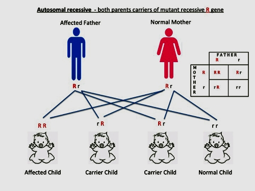
  • If both parents are carriers of the mutant allele, the probability is 25% chance of affected children (homozygous), 50% of children have chance of carriers and 25% chance of children having no mutant allele.
 Examples:
  • Sickle cell disease
  • Cartilage hypoplasia
  • Congenital insensitivity to pain
  • Diastrophic dwarfism
  • Gaucher disease
  • Hurler syndrome
  • Hypophosphatasia
  • Manteaux syndrome
  • Alkaptonuria

BS8GENETICS4.jpg

Figure 4. Schematic diagram and Punnett Square showing inheritance pattern in autosomal recessive genetic disorder.

  • Males and females have the condition in roughly equal proportions.
  • Patients with the condition are usually in one sibship in one generation.
  • Affected offspring have normal parents.
  • Consanguinity, where both parents have one or more ancestors in common, increases the chance that a condition presenting in a child of theirs might be due to both parents being carriers for the same recessive gene alteration.

BS8GENETICS5.png

Figure 5. Pedigree of AR inheritance

Sickle cell disease

  • A group of disorders caused by a mutation in the beta globin gene (HBB).
  • The altered haemoglobin produced is HbS.
  • SCA is the commonest of these diseases and is caused by homozygous point mutations in the HBB gene on the short arm of chromosome 11.
  • A single nucleotide mutation (base change) from T to A results in glutamic acid changing to valine at the 6th amino acid in the beta haemoglobin chain (Glu6Val or E6V)
  • This causes the abnormal haemoglobin S subunits to stick together and form long, rigid molecules that bend red blood cells into a sickle (crescent) shape.
  • The sickle-shaped cells die prematurely, which can lead to anaemia. The sickle-shaped cells are rigid and can block small blood vessels, causing severe pain and organ damage.

Clinical features

  • SCA is characterised by episodes of pain owing to vaso-occlusive events, chronic haemolytic anaemia and severe infections from early childhood with splenomegaly.
  • Any organ may be affected but most commonly bones (AVN/osteomyelitis), lungs, liver, kidneys, brain, and eyes are involved.
  • Individuals can clinically manifest when only one copy of the mutant allele is present.
  • There is no transmission from affected father to son. There can be transmission from father to daughter – all daughters of an affected male will inherit the condition.
  • Affected mother can cause transmission to 50% of sons and daughters.

Example:

  • Hypophosphatemic rickets.

BS8GENETICS6.jpg

Figure 6. Schematic diagram and Punnett Square showing inheritance pattern in X-linked dominant genetic disorder 

  • The key for determining if a dominant trait is X-linked or autosomal is to look at the offspring of the mating of an affected male and a normal female.
  • If the affected male has an affected son, then the disease is not X-linked. All of his daughters must also be affected if the disease is X-linked.

BS8GENETICS7.png

Figure 7. Pedigree of X linked dominance inheritance

  • X-linked recessive traits are fully manifest in males because they only have one copy of the X chromosome.
  • Females are usually affected when both copies of X chromosomes have the mutant allele (homozygous).
  • Males are more commonly affected than females for the above reasons.
  • If the mother is a carrier – 50% of daughters and 50% of sons inherit the mutant allele.
  • If the father has the mutant allele – there is no transmission to sons but all daughters will inherit the mutant allele (carriers).
  • If both father and mother have the mutant allele –
  • Examples:
    • Duchenne’s muscular dystrophy
    • Haemophilia A
    • Hunter syndrome

BS8GENETICS8.jpg

Figure 8. Schematic diagram and Punnett Square showing inheritance pattern in X-linked recessive genetic disorder 

  • Trait skips generations.
  • Affected fathers DO NOT pass to their sons.
  • Males are either affected or normal, never carriers.
  • All affected males in a family are related through their mothers.
  • Trait or disease is typically passed from an affected grandfather, through his carrier daughters, to half of his grandsons.
  • Males are much more likely to be affected than females. If affected males cannot reproduce, only males will be affected.
  • Carrier females may show variable expression due to lyonisation (X inactivation).
  • Females only affected if they get a copy from both parents.

BS8GENETICS9.jpg

Figure 9. Pedigree of X linked recessive inheritance

Duchenne’s muscular dystrophy

  • Muscular Dystrophy (DMD) is one of the dystrophinopathies caused by a mutation in the dystrophin gene (Xp21).
  • The dystrophin protein provides structural stability to the dystroglycan complex of the muscle cell membrane, which is lost as a result of the mutation
  • Blastocyst: The 4–9-day-old embryo (post-fertilisation) which consists of 100–200 total cells. This stage of development is prior to implantation in the uterus. Only two types of cells are present at this time, the trophectoderm (foundation of the placenta) and the inner-cell mass, which will also contribute cells to the extraembryonic tissues as well as the entire fetus.
  • Inner cell mass (ICM): A population of cells (approximately 20–30%) at the blastocyst stage that generates certain extraembryonic cells and tissues as well as those of the entire embryo.
  • Epigenetic: Chemical modifications of DNA that do not alter a gene’s sequence, but impact gene expression.
  • Genome: The entire set of genetic material in an organism.
  • Genotype: The DNA sequence of a gene.
Previous
Next