TOPIC DETAILS

  Team Member Role(s) Profile
Paul Banaszkiewicz Paul Banaszkiewicz Section Editor
Terence Terence Savaridas Segment Author

Bone mass relates to the composition of cells, minerals and architecture of a given volume of bone. 

Bone mass defines the material properties of bone. This influences the load to failure of bone and therefore is a factor in assessing fracture risk. 

Bone mass is frequently assessed using radiological parameters

Peak bone mass is estimated to occur between the ages of 16 to 25 years.  This is influenced by multiple factors:

i.                Genetic – racial variations, rickets, hypophosphatasia.

ii.              Gender – greater in males.

iii.             Nutritional status – malnutrition, malabsorption.

iv.             Endocrinopathy – PTH overproduction (MEN, pituitary adenoma).

v.              Iatrogenic – anticonvulsant medication (phenytoin)

vi.            Load bearing – bone modeling occurs in response to load. In the absence of load bearing e.g. paresis peak bone mass attained will be diminished.

Age and bone mass (Figure 1)

Once peak bone mass is obtained, in the 3rd decade, there is a complex interaction between bone deposition and bone resorption in order to maintain an adequate bone mass.

In spite of this it is estimated that the average adult sustains an average bone loss of 0.3% - 0.5% per year.

In females, following menopause the estimated rate of annual bone loss is tenfold greater at approximately 3% per year.

This, in addition to the lower peak bone mass attained in females contributes to the greater fracture risk seen in post-menopausal women.

On a cellular level the regulation of bone mass is mainly dependent on the function of osteoclasts and osteoblasts.

-        Osteoclast: Essential for bone resorption.

-        Osteoblast: Bone forming cell.

The function of these two cell types are coupled to each other.

BS9BONEMASS1a.png

Figure 1. Age and bone mass

Mechanism of Action

1.      Osteoclast

-        Attracted to areas of ‘micro-damage’ where its ruffled border attaches to create a sealed cavity.

·       Via an energy dependant process carbonic acid and matrix degrading enzymes are secreted in to this sealed cavity causing bone resorption.

-        Following bone resorption by osteoclast, osteoblast lay down osteoid that subsequently forms new bone.

·       Osteoclast are stimulated by RANKL which is produced both by osteoblast and osteocytes.

2.      Osteoblast

-        Osteoblast activity is influenced by osteocytes.

3.      Osteocytes

·       The most abundant cell type in bone. They form an interconnected network in mineralised bone.

·       They respond to mechanical changes applied to bone and are often referred to as the ‘mechanostat’.

o    Respond to mechanical change on bone. 

-        Secretes

·       Sclerostin that has an inhibitory effect on bone formation by blocking osteoblast function. When bone is loaded, there is a decrease in sclerostin secretion and this results in increased bone formation by osteoblast via the Wnt pathway. 

·       RANKL secretion, in contrast, is increased when bone is unloaded. This stimulates osteoclastic mediated bone resorption.
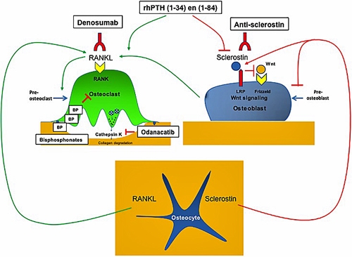
BS9BONEMASS2.jpg 

Figure 2. Diagram demonstrating  the effect of antiresorptive and osteoanabolic drugs on bone.

1.      Clinical Practice

2.      Research

1. Clinical Practice

 a) DEXA (Dual-Energy X-ray absorptiometry) Bone Scan

-  This remains the gold standard evaluation of bone mass in clinical practice.

- The energy of an X-ray beam that is passed through bone is absorbed and the remainder is detected on the other side of the body. In bone of greater density, more energy is absorbed.

- In DEXA scanning, X-ray beams of two different energies are used. This allows calibration to subtract the energy that is absorbed by fat and soft tissues, hence providing a truer estimate of bone density.

- DEXA values for bone mass are quoted as g/cm2 or more frequently converted to values that relate to the average peak bone mass (T-score) or the average for a given patients’ age (Z-Score).

  • T-Score: Bone density measurement in relation to the peak bone mass of healthy young adult of similar gender.
  • Z-Score: Bone density measurement in relation to the average bone mass of an adult of similar age and gender.

- Measurements are frequently taken of the:

  • Lumbar spine: L3, L4
  • Proximal femur

-  The WHO (World Health Organisation) guideline for interpretation of DEXA results is based on T-score measurements;

  • Normal: Score not more that 1 standard deviation (SD) below the average young adult value.
  • Osteopenia: Score between 1 and 2.5 SD below that of the adult average.
  • Osteoporosis: Score of <2.5SD below the average value for a young adult.
  • Severe osteopososis: Score of <2.5SD  and with a history of fracture.

BS9BONEMASS3.png

Figure 3. In bone mineral density (BMD) scanning, the energy of x ray beams that are passed through bones is absorbed, and what is not absorbed is detected on the other side of the body. The more dense the bones (from greater mineral content), the more energy is absorbed, and the less energy detected.

Previous
Next