TOPIC DETAILS

  Team Member Role(s) Profile
Paul Banaszkiewicz Paul Banaszkiewicz Section Editor, Segment Author
Chris Ghazala Christopher George Ghazala Segment Author
  • Meniscus is a definite A-list topic that will be tested in both the basic science and pathology viva sections.
  • An understanding of the normal anatomy, physiology and biomechanics of the menisci is a necessary prerequisite to understanding the pathogenesis of disorders involving the knee.
  • Elastofibrocartilagenous.
  • Crescent shaped; triangular appearance on cross-section.
  • Anterior horns attached to each other by the small transverse anterior intermeniscal ligament.
  • The medial meniscus (MM) covers 64% of the condylar surface and is more C-shaped.
  • The MM is asymmetrical, having a considerably wider posterior than anterior horn.
  • The MM has firm attachments all around its periphery via the coronary (meniscotibial) ligaments and attaches to the deep part of the medial collateral ligament.
  • Its anterior attachment is approximately 10 mm anterior to the anterior cruciate ligament (ACL) insertion in line with the medial tibial tubercle.
  • Posteriorly the MM is attached to the posterior intercondylar fossa of the tibia directly anterior to the posterior cruciate ligament (PCL) attachment.
  • The lateral meniscus (LM) is more circular and covers a larger area of the condylar surface (84%).
  • The LM has anterior and posterior horns the same width.
  • The LM has twice the excursion of the medial meniscus during knee motion.
  • Anterior horn of lateral meniscus and post horns of both meniscii attach to the intercondylar eminence.
  • Popliteus muscle (not tendon) is attached to lateral meniscus.
  • Semimembranosis is attached to medial meniscus.
  • The lateral meniscus is connected to the femur either in front of (anterior meniscofemoral ligament of Humphrey) or behind (posterior meniscofemoral ligament of Wrisberg) the PCL.

BS2M 1.jpg

Figure 1. Labelled axial view of a knee showing sections of the meniscus and their relationships

BS2M 10.png

Ultrastructure of meniscus 

Water

  • The menisci are composed of 65–75% water.

Collagen

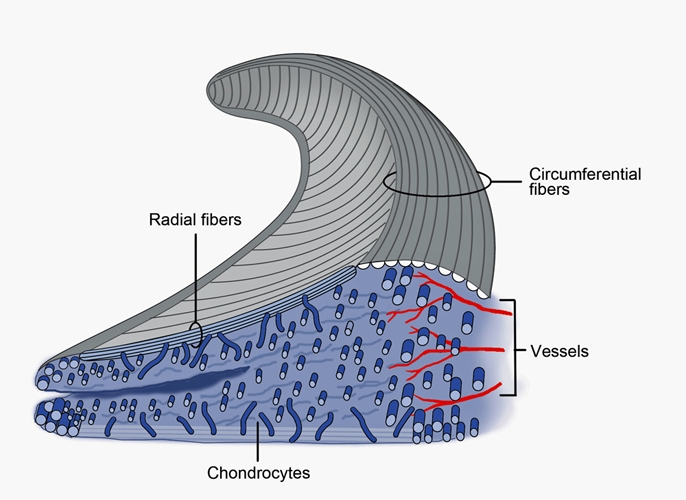
  • There are three layers of collagen fibres: superficial, lamellar and deep.
  • The majority of collagen fibres are arranged longitudinally (circumferential) in the deep layer.

Proteoglycans

  • Located within a fine meshwork of collagen fibrils, proteoglycans are large, negatively charged hydrophilic molecules.
  • Aggrecan is the major large proteoglycan of the meniscus while biglycan and decorin are the main small proteoglycans.
  • Their main function is to enable the meniscus to absorb water, whose confinement supports the tissue under compression.

Elastin

Glycoprotein

  • Fibrochondrocytes that synthesis and maintain the extracellular matrix. There are two types of cells:
  • Fusiform cells found in the superficial zone
  • Ovoid cells found elsewhere
  • The outer proportion of the meniscus contains mainly type 1 collagen compared to the inner portion that contains 60% type II and 40% type I 
  • Therefore the outer proportion of the meniscus is more fibrous and withstands tensile stress but has low compressive and torsional stiffness. The inner portion is able to withstand compression better.
  • The menisci are microscopically arranged into three distinct layers: superficial, lamellar and deep.
BS2M 2.jpg

Figure 2. A cross-section of the meniscus showing the radial and circumferential collagen fiber orientation. Numerous bundles of circumferentially oriented Type I collagen fibers are dispersed throughout the meniscus giving its highest tensile strength in that direction. Other collagen fibers are oriented radially and woven into the circumferential bundles. There are also some superficial fibers with random orientation creating a mesh-like matrix on the femoral articular surface.

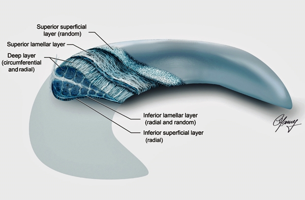
  • The superficial layer exists on both the tibial and femoral surfaces. It is composed of an unorganised random arrangement of collagen fibrils in the superior region and more radially orientated fibres in the inferior region. This surface arrangement allows the meniscus to slide optimally along the articulating surfaces of both the femoral condyles and tibial plateau.
  • Underneath the reticular is a lamellar layer that also exists on both the femoral and tibial sides. These fibres are variable in direction with a predominantly radial orientation at the periphery and at the anterior and posterior horns. Despite the presence of radial fibres, random fibre orientation dominates the lamellar zone.
  • The main part of the meniscal tissue is located between the two lamellar layers. A dense framework of circumferential coarse type I collagen fibreslies in this layer, cross-linked with radial fibres from the periphery. The radial fibres may act as a “tie” holding the circumferential fibres together, providing structural rigidity against compressive forces and resisting longitudinal splitting of the menisci.
BS2M 3.jpg

Figure 3. Collagen fiber arrangement in the meniscus. Collagen organisation should be considered as 3 layers: superficial, lamellar and deep. Collagen fibers are unstructured in the superior superficial layer but radially orientated in the inferior superficial layer. Amorphous collagen arrangement persists in the lamellar layer but is different from the superficial layer in that contains short, radially orientated fibers only at the posterior and anterior horns.

  • The development of “hoop stress” within the meniscus depends on intact anterior and posterior attachments. Hoop stress also relies on the conversion of axial load into tensile strain through intact longitudinally oriented collagen fibres.
  • The compression of the menisci by the tibia and femur generates outward forces that push the meniscus out from between the bones.
  • The circumferential tension in the menisci counteracts this radial force.
  • Hoop stress is the stress in a direction perpendicular to the axis of an item.
  • As the thickness of the item decreases the hoop stress increases.

BS2M 4.jpg

Figure 4. The importance of intact meniscal entheses for the load distribution function of the meniscus. With intact enthesis the load  is transmitted via the menisci and articular cartilage through a large contact area.Part of the load is transformed to hoop stresses.
 
BS2M 5 .jpg

Figure 5. Hoop stresses on the meniscus

BS2M 6.jpg
 
Figure 6. Hoop stresses
  • The primary functions of the menisci are load transmission and shock absorption. The distributed joint force is applied normal to the surface of the menisci.
  • Due to the wedge shape of the meniscus, this normal force can be divided into an axially orientated compressive force component and a radially orientated force component.
  • The circumferentially orientated fibre bundles are stiff and resist the radially directed force from the femur with tension force that travels circumferentially through the meniscus and into the attaching ligaments. The circumferential force in the meniscus produces hoop strain and hoop stress.
  • Menisci are always compared to the hoops around a wooden wine barrel – because of the arrangement of those hoops the barrel is extremely strong, but when you take one of those hoops away it becomes weak. If the meniscus is damaged and torn in a longitudinal fashion it follows the hoops, and it is not nearly as bad as if you tear it in a radial fashion when you tear across those hoops and completely de-function the meniscus.

BS2M 7.jpg

Figure 7. As the femur presses down on the meniscus during normal loadiing,the meniscus deforms radially but is anchored by its anterior and posterior horns(Fant and Fpost)During loading tensile,compressive and shear forces are generated.A tensile hoop stress(Fcir) results from radial deformation,while vertical(FV) and horizontal forces(Fh) result from the femur pressing on the curved superior surface of the tissue.A radial reaction force(Frad) balances the femoral horizontal force(Fh)

  • Branches of the lateral, middle and medial genicular arteries from the popliteal artery provide the major vascularisation to the inferior and superior aspects of each meniscus.
  • The middle geniculate artery is a small posterior branch that pierces the oblique popliteal ligament at the posteromedial corner of the tibiofemoral joint.
  • A premeniscal capillary network arising from branches of these arteries originates within the synovial and capsular tissues of the knee along the periphery of the menisci.
  • Only 10–30% of the peripheral medial meniscus border and 10–25% of the lateral meniscus border receives direct blood supply.
  • The inner two-thirds of the meniscus is avascular and is nourished by the synovial fluid through diffusion.
  • Although fully vascularised at birth, the blood vessels in the meniscus recede during maturity. In adulthood, the red–red region contains the overwhelming majority of blood vessels.

BS2M 8 .jpg

Figure 8. Frontal section of the medial compartment demonstrates the microvasculature of the medial meniscus. The perimeniscal capillary plexus (PCP) permeates through the peripheral border of the meniscus

Load bearing

  • At least 50% of the compressive load of the knee joint is transmitted through the meniscus in extension, and approximately 85% of the load is transmitted in 90 degree flexion.
  • In the meniscectomised knee the contact area is reduced to approximately 50%.
  • Partial meniscectomy also increases the contact pressures.

Shock absorption

  • Menisci may attenuate the intermittent shock waves generated by impulse loading during gait – the shock absorbing capacity of normal knees is approximately 20% higher than in meniscectomised knees.
  • The inability of a system to absorb shock has been implicated in the development of osteoarthritis.1

Articular conformity

  • The meniscus displaces in an anteroposterior direction as the knee passes through its range of movement.
  • In addition, the menisci deform to remain in constant congruity to the tibial and femoral articular cartilage.
  • Deformable properties of the meniscus aids load transmission and shock absorption.

Knee joint stability

  • The medial meniscus in particular controls anteroposterior translation.
  • Meniscectomy alone may not seriously affect stability. However, in association with ACL tears, meniscectomy significantly increases the anterior knee laxity.

Lubrication

  • Distributes synovial fluid and promotes a viscous hydrodynamic action needed for fluid film lubrication.

Proprioception

  • This has been inferred from the finding of type 1 and type 2 nerve endings in the anterior and posterior horns of the menisci.

Prevention of soft tissue impingement during joint motion

Biomechanics 

  • The lateral meniscus provides more biomechanical support to the joint than does the medial meniscus.
  • Partial meniscectomy causes increased contact pressures on the articular cartilage.
  • Classified either on location in respect to blood supply or tear pattern.
  • Meniscal tears can be either traumatic or degenerative in nature.
  • Meniscal tears are uncommon in persons under 10 years of age, but become increasingly common during and after adolescence.
  • Degenerative tears can be found in as much as 60% of the population over the age of 65 years. The majority of these tears, however, are asymptomatic and occur in association with degenerative joint disease. The changing patterns of meniscal injury with chronological age most likely correlate with normal alterations in collagen fibre orientation with aging, as well as increasing intrasubstance degeneration.
  • The majority of meniscal tears affect the medial meniscus and tend to involve the posterior horn.
  • Meniscal tears are either partial or full thickness and stable or unstable.
  • An unstable tear is one in which the entire tear or a portion thereof can be displaced into the joint space. There it may become trapped, causing pain by traction at the meniscocapsular junction. It may be responsible for symptoms of catching, locking and effusion.
  • Meniscal injuries can be further classified based on their tear patterns:
  • Vertical
  • Horizontal
  • Complex
  • The circumferential continuity of the peripheral rim of the meniscus is integral to meniscal function. Partial meniscectomy, or bucket-handle tearing, will still preserve meniscal function so long as the peripheral rim is intact. However, if a radial tear extends to the periphery and interrupts the continuity of the meniscus, the load-transmitting properties of the meniscus are lost.
  • Under axial femoral compressive loads, the peak contact stress and maximum shear stress in the articular cartilage increased 200% more after a lateral than a medial meniscectomy. These increased stresses may explain the higher cartilage degeneration observed after a lateral meniscectomy.
BS2M 9.jpg
Figure 9. Types of meniscal tear

Open repair

  • Advantage of better preparation of the tear site.
  • Only the most peripheral of tears in the anterior aspect of the knee involving the red–red zone are amenable to this technique because of exposure and accessibility.
  • Due to improved arthroscopic techniques rarely performed these days.

Arthroscopically assisted

  • Inside-out technique:
  • First described by Henning.
  • Utilises zone-specific cannulas to pass sutures through the joint and across the tear. The sutures are swaged onto flexible needles. A small posterior joint line incision is used to retrieve the sutures and tie directly on the capsule. The use of a posterior retractor, such as a gynaecological speculum, is vital in order to protect the posterior neurovascular structures.
  • Outside-in techniques:
  • Described by Warren and Morgan and Casscells.
  • Involve passing sutures percutaneously through spinal needles at the joint line across the tear, and then retrieving the sutures intra-articularly. Mulberry knots can then be tied on the intra-articular free ends of the suture. A small incision is then made at the joint line,where the protruding suture ends are retrieved and tied directly on the capsule. An alternative technique is to retrieve the intra-articular portion of the suture with another pass across the tear using a wire snare and tying the suture back on itself on the capsule. This technique eliminates the need for Mulberry knots.
  • This technique is mainly only used for longitudinal tears particularly those of the anterior horns, where arthroscopic assess is difficult.
  • A potential disadvantage of the outside-in technique is difficulty in reducing the tear and opposing the edges while passing the sutures.
  • All-inside technique:
  • Suitable for repairs of the far posterior horns.
  • Use has increased significantly since the introduction of implantable anchors, arrows, screws, and staples.
  • Although the pullout strength of some of these devices has been shown to approximate those of mattress sutures in cadaveric studies, there have been no long-term clinical studies that compare them to more traditional repair techniques.

Healing

  • Similar to other connective tissues – exudation, organisation, vascularisation, cellular proliferation, remodelling.
  • Following injury there is formation of a fibrin clot rich in inflammatory cells. Vessels from the perimeniscal capillary plexus proliferate into this fibrin scaffold, followed by mesenchymal cell proliferation forming a cellular fibrovascular scar. Modulation of this scar tissue into normal-appearing fibrocartilage requires several months.
  • Approximately 80% of repairable menisci are found in knees with an acute or chronic tear of the ACL – thus repair of the meniscus is linked to the management of the ACL tear.
  • NB. Risk of injury to peroneal nerve in lateral meniscus repair, saphenous nerve in medial repair.
  • Three zones determine the healing prognosis for meniscal lesions: red–red, red–white and white–white:
  • The red–red zone is fully vascular and therefore has an excellent healing prognosis.
  • The red–white zone is at the border of vascular supply and has a generally good healing prognosis.
  • The white–white zone is relatively avascular and has a poor prognosis for healing.
  • Cadaveric menisci are matched by site and size, and are implanted using various methods that include a free soft tissue allograft implantation, separate anterior and posterior bone plugs.
  • The criteria for meniscal transplant include:
  • Younger than 55 years and physically active.
  • Missing more than half of a meniscus as a result of previous surgery or injury, or a meniscus tear that cannot be repaired.
  • Persistent activity-related pain.
  • Knee with stable ligaments and normal alignment.
  • No or minimal knee arthritis.
  • Not obese.
  • These cells can differentiate into chondroblasts and help in the meniscal repair process.
Previous
Next

QUESTION 1 OF 1

A 44-year-old male attends the Outpatient cliniic complaining of a 3 months history of knee pain, swelling and stiffness.His MRI scan is shown below(Figure 1.1)
Concerning his symptoms

MTI.jpg 


QUESTION ID: 1048

1. Clinical diagnosis is usually straightforward
2. On the affected side(lateral) the tibiofemoral contact pressure not significantly increased
3. Partial meniscectomy partially restores native joint biomechanics
4. Results in an increased tibiofemoral contact pressure equal to around 70% of that following a total meniscectomy
5. There is usually a history of an acute traumatic event

Further Reading

  • 1. McDermott ID. Meniscal tears. Curr Orthopaed 2006; 20: 85–94.
  • 2. Strauss EJ, Day MS, Ryan M, Jazrawi L. Evaluation, treatment and outcomes of meniscal root tears. JBJS Rev 2016; 4(8): e2 1–10.

References

  • 1. Radin EL, Rose RM. Role of subchondral bone in the initiation and progression of cartilage damage. Clin Orthop Relate Res 1986, 213: 34–40.