TOPIC DETAILS

  Team Member Role(s) Profile
Paul Banaszkiewicz Paul Banaszkiewicz Section Editor
Hussein H Noureddine Segment Author

For the average orthopaedic surgeon embryology is a daunting complicated subject to learn. However, a basic understanding of skeletal embryology is essential especially in correlation to spinal cord and limb formation as a multitude of pathologies are related to anomalies arising during the embryological stages.

For detailed embryological information, the reader should study the Carnegie series at:

http://php.med.unsw.edu.au/embryology/index.php? title=Embryonic_Development.

BSEMBRYOLOGY 1.png

Figure 1.Human embryo.Image source:Pooh et al. Human embryo MR imaging microscopy and high-resolution transvaginal 3D sonography. Volume 204, Issue 1, January 2011, Pages 77.e1–77.e16

Carnegie stages are a system used by embryologists to describe the apparent maturity of embryos. An embryo is assigned a Carnegie stage (numbered from 1 to 23) based on its external features. This staging system is not dependent on the chronological age nor the size of the embryo. The stages are, in a sense, arbitrary levels of maturity based on multiple physical features. Embryos that might have different ages or sizes can be assigned the same Carnegie stage based on their external appearance because of the natural variation which occurs between individuals.

  • The period of time from fertilisation to birth is divided into three periods:
  • Pre-embryonic period from fertilisation to week 3
  • Embryonic period from weeks 3 to 9
  • Fetal period from 9th week until birth
  • Formation of trilaminar embryo.

Week 3

  • Gastrulation is the formation of the trilaminar embryonic disc from a bilaminar disc. Epiblast becomes ectoderm (dorsal), hypoblast becomes endoderm (volar)
  • The primitive streak at the dorsal aspect of the embryonic disc is formed. The cranial end thickens to form the primitive knot, defining the cranial end. (Primitive streak remnants can form teratomas.)
  • The primitive streak produces mesenchymal (mesoblastic) cells destined to form the mesoderm by blending endoderm and ectoderm. This will produce connective tissue, bone, blood vessels, blood cells, muscles and the genitourinary system.
  • Ectoderm forms the notochord - at the cranial end of the primitive streak. The notochord is the structure around which the spinal cord will develop (remnants can form chordomas).

BSEMBRYOLOGY 2a.pngBSEMBRYOLOGY 2b.png

Figure 2. Carnegie stage 10 embryo Three-dimensional computer graphics model of Carnegie stage 10 embryo with closing neural tube. Ten pairs of somites are recognizable. Round ball-like structure on ventral side of embryo is yolk sac. Model was reconstructed based on its gross photographs and histologic sections. Image source: Pooh et al. Human embryo MR imaging microscopy and high-resolution transvaginal 3D sonography. Volume 204, Issue 1, January 2011, Pages 77.e1–77. e16

Neurulation

  • The notochord induces the overlying ectoderm to form the neural plate (the primordium of the central nervous system (CNS)).
  • The thickened plate then invaginates centrally to form the neural groove, with thickened neural folds (day 18). The folds fuse dorsally forming the neural tube, which will become the spinal cord.
  • As the neural folds fuse some of the ectodermal cells lying along the crest of each neural fold attach to neighbouring cells and become the neural crest cells, which differentiate to begin the formation of the peripheral nervous system, the automatic nervous system and Schwann cells.

Somatisation

  • Somites are formed from mesoderm and they begin to line both sides of the notochord. Eventually they will form 42–44 pairs. The somites continue their developmental process and soon become a lateral dermatome, a medial myotome and a ventral scleratome. This becomes, in due time, the basis of the skin, muscle and skeletal elements, respectively.

BSEMBRYOLOGY 3a.pngBSEMBRYOLOGY 3b.png

Figure 3. Carnegie Early Stage 10 (22 - 23 days) 2 - 3.5 mm This is a dorsal view of the embryo. Amniotic membrane removed. Somite Number 4 - 12, rostral neuropore, neural folds in region of developing brain, neural tube, somites, caudal neuropore, neural fold fuses, remnant of amniotic sac. Image source: The Kyoto Collection images are reproduced with the permission of Prof. Kohei Shiota, Anatomy and Developmental Biology Kyoto University Graduate School of Medicine, Kyoto, Japan for educational purposes only.

Weeks 4 to 6

  • Main organs laid down resulting in a miniature humanoid form 3 cm long. Complexity of tissue interactions explains susceptibility to environmental exposures (“critical period”).
  • The embryo folds laterally and transversally forming a C-shaped cylindrical embryo.
  • The apical ectodermal ridge (AER) exerts an inductive influence on the limb mesenchyme that promotes growth and development of the limbs.
  • Upper limbs: at this time the limb buds also develop. The upper extremity, with pronated forearms, appears first, and begins to rotate externally.
  • Lower limbs: the lower extremity appears slightly later than the upper extremity and begins to rotate internally.
  • Developmental disturbances during this period give rise to major congenital malformations. Early suppression of limb development causes amelia (complete absence of a limb); late suppression causes meromelia (partial absence).

BSEMBRYOLOGY4.png

Figure 4.Stage 13 Week 4-5, 26 - 30 days, 3 - 5 mm, Ectoderm: Neural tube continues to close, Caudal neuropore closes, forebrain. Mesoderm: continued segmentation of paraxial mesoderm (21 - 29 somite pairs), heart prominence .Head: 1st, 2nd and 3rd pharyngeal arch, forebrain, site of lens placode, site of otic placode, stomodeum. Body: heart, liver, umbilical, early upper limb bulge Image source Dr Steven O'Connor (Houston, Texas) from http://php.med.unsw.edu.au/embryology/index.php?title=File:Stage13_bf5.gif

Week 7 to birth

  • Maturation with major emphasis on bone and CNS development. Thus growth and mental function affected by environmental insults.
  • By week 7 the 10 finger rays appear and continue to differentiate until weeks 12–13 when the hands appear.

BSEMBRYOLOGY 5a.pngBSEMBRYOLOGY 5b.png

Figure 5.Human foetus Carnegie stage 20 – 8 weeks.The cartilaginous precursors (anlagen) of the long bones appear early in embryonic life

  • During this initial 12-week period the formation of the body’s solid framework also begins. The beginning process involves mesenchymal aggregation into a cartilage prototype. Gradually but systematically each cartilage model becomes solid bone. This process applies to all bones except those formed through intramembranous ossification such as the skull.
  • By week 12 the primary centres of ossification in the diaphyses of most bones have appeared.

BSEMBRYOLOGY 6a.pngBSEMBRYOLOGY 6b.png

Figure 6.The cartilaginous anlage is invaded in its central segment by blood vessels.

  • Gradually but systematically each cartilage model becomes solid bone. This process applies to all bones except those formed through intramembranous ossification such as the skull.
  • This process starts with the invasion of the centre of the shaft of the cartilaginous anlage by blood vessels. This leads to endochondral ossification – the histological specimen is of a fetal rat’s tibia.

BSEMBRYOLOGY 7a.pngBSEMBRYOLOGY 7b.png

Figure 7. Tibia Shaft Ossification(Histology specimen young rats tibia)

  • This process results in centripetal ossification of the cartilaginous anlage’s shaft, producing the early diaphyseal bone.

BSEMBRYOLOGY 8.png

Figure 8.Primary ossification centres diaphyses 

  • By week 12 the primary centres of ossification in the diaphyses of most bones have appeared.

BSEMBRYOLOGY 9a.pngBSEMBRYOLOGY 9b.png

Figure 9.Invasion of the cartilaginous end caps (epiphyses) by blood vessels, resulting in the formation of the epiphyseal ossific nuclei (secondary ossification centers).

  • Postnatally, the next skeletal event is the invasion of the cartilaginous end caps (epiphyses) by blood vessels, resulting in the formation of the epiphyseal ossific nuclei (secondary ossification centres).
  • The intervening cartilaginous disc between the primary and secondary ossification zones forms the future growth plate (physis).
  • The Homeobox genes control mass and local growth in a distal direction.
  • The AER releases a factor that diffuses proximally for a distance of 300 microns. Cells within this distance of the AER are lying within the progress zone. Initially a limb bud is small and all of the cells lie in the progress zone.
  • They have a low value. Each time a cell divides in the progress zone, it gains a higher value. But cell division also leads to growth of the limb bud, and some cells now lie outside the progress zone.
  • These cells can divide but they no longer increase in value, instead they differentiate with their value dictating the structure they differentiate into.
  • The zone of polarising activity (ZPA) is a specialised group of cells on the caudal edge of the limb bud.
  • The ZPA under control of Sonic Hedgehog gene is a source of a diffusible substance or morphogen, which establishes a gradient across the limb bud.
  • Complex cell-signalling possibly with vitamin A (retinoic acid) being involved, results in gradual switching off of HOX genes and cells differentiate into relevant digits.
  • Vertebral formation at 3–5 weeks with segmentation occurring at 6–8 weeks.
  • Each vertebrae forms from two adjacent sclerotomes and so becomes an inter segment structure.
  • Notochord degenerates but between vertebrae it persists to form the nucleus pulposus.
  • During the 6th week chondrification occurs.
  • Two centres in each centrum fuse at the end of the embryonic period resulting in one centre evident in the centrum after this period (defects here ®hemi vertebrae).
  • Centres in the neural arches fuse with each other and the centrum.
  • The two halves of the neural arch usually fuse in the first year and these to the centrum in approximately the 3rd to 6th year.
  • At about puberty five secondary centres appear (upper and lower body, and one in the tip of the transverse processes and spinous process).

BSEMBRYOLOGY10c.png

Figure 11.Spinal development controlled by Homeobox gene

  • Ossification begins in the embryonic period and ends at about 25 years.
  • Longitudinal growth is by way of superior and inferior apophysis.
  • Horizontal growth is by periosteal apposition.
  • Spinal canal enlarges by growth of the pedicles and posterior elements, enlarging rapidly from birth to 5 years and more slowly from 5 to 10 years.
  • The spinal canal reaches its final dimensions relatively early compared with the continual growth of the rest of the vertebral structures.
  • The final height of vertebral column is reached in girls by 11–13 years and in boys by 14–16 years.

CASE BASED DISCUSSIONS

Case based Discusson Limb Bud

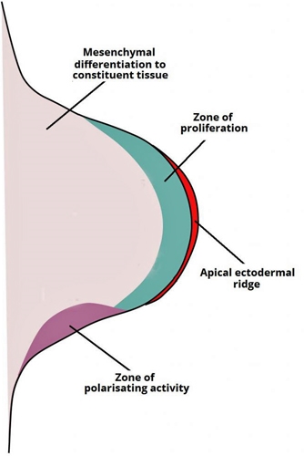
Knowledge of the embryonic limb bud and its different zones and is essential to understand congenital limb deformities. The limb bud is made of mesoderm covered by ectoderm, and the upper limb bud appears toward the end of the third week followed by the lower limb bud which appears about 4 days later (fourth week).

The Apical Ectodermal ridge is the most distal zone of the bud (made of the covering ectoderm) and controls proximal to distal growth or elongation. Deep to the AER is the progress zone that is made of rapidly dividing mesoderm cells which enables this longitudinal growth. AER is controlled by the FGF gene family and injuries to AER will lead to congenital amputation, defects in the ridge can lead to truncation (such as club hand).

The AER appears first then the Zone of Polarizing Activity (ZPA) appears along the posterior aspect of the limb bud and signals the anteroposterior (radio-ulnar) growth. It is under the control of the SHH compound. Abnormalities in the ZPA can lead to a range of abnormalities from congenital absence of thumb (anterior / radial abnormalities) to ulnar polydactyly with SHH upregulation to loss of ulnar digits in the case of SHH downregulation (posterior /ulnar abnormalities).

The third signalling centre controls dorso-ventral growth comprises the rest of the ectoderm (the non AER area of ectoderm) and is controlled by the WNT gene.
 BSE.jpg

Figure 11.Limb elongation occurs through proliferation of the underlying mesenchyme core, in which the AER plays a crucial role in ensuring that the mesenchyme immediately underneath it remains undifferentiated. As growth proceeds, the proximal mesenchyme loses signals from the AER and begins to differentiate into the constituent tissues of the limbs.

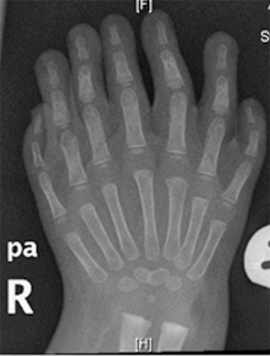
Examiner: Please have a look at this X ray and describe what you can see.(Figure 11)
BSE 11.png

Figure 12.Radiograph hand

Candidate: There is symmetric polydactyly with absence of radial rays, these are usually features of ulnar dimelia or mirror hand which is a congenital condition.

Examiner: That is correct, what is the genetic defect that would lead to this

Candidate: Sonic hedgehog (SHH) gene, which controls the Zone of polarizing activity of the limb bud which in turn signals the Antero-posterior or radio-ulnar limb growth.

Examiner: If Upregulation of the SHH gene leads to ulnar duplication why do we see associated radial absence

Candidate: The posterior or ulnar components form earlier than the anterior or radial components so a disruption the antero-posterior signalling leading to an ulnar duplication due to upregulation of the SHH will also lead to losing the elements that form later ie the radial ones.

Case based discussion:Spinal cord embryology

Basics and Definitions: In the early embryonic stages there are three primary cellular layers (also known as germ layers), the outer is the ectoderm and gives the epidermis and the neuro-ectoderm (which includes the neural crest and neural tube). The middle is the mesoderm which mostly forms muscles, cartilage, and bone. And the endoderm is the inner layer which forms columnar cells lining multiple gastro-intestinal organs.

Neural Tube: Results from the process of neurulation of the ectoderm where the latter divides into a neural tube in the inside, an epidermis on the outside, and neural crest cells in between which then migrate to other locations. The neural tube starts as a flat neural plate the edges of which thicken and fold closing over to form the tube. It eventually develops into four subdivisions; the forebrain, midbrain, hindbrain, and spinal cord. As the closure progresses through folding the open ends of both sides of the tube are called the neuropores with anterior (cranial) neuropore and posterior (caudal) neuropore. These should close by the end of the week 4 and failure to close leads to Neural Tube Defects Which are anencephaly for the anterior / cranial neuropore and spinal dysraphism for posterior / caudal neuropore.

BSEMBRYOLOGY 10b.png

Figure 13.Spinal cord embryology

Notochord: is a flexible rod like structure derived from mesoderm that provides support to the embryo. It just precedes neurulation and ends up being situated between the neural tube which is dorsal to it and the endoderm layer which is ventral to it. The Notochord is separated from the neural tube by further mesoderm that is comprised of somites (mesoderm repeating blocks). The somites give rise to dermatomes, myotomes and sclerotomes. The sclerotomes form as paired condensations around the notochord. Extensions from each postero-lateral half of the condensation extend dorsally around the neural tube and meet posterior to it.  The Sclerotomes split in the middle (Von Ebner Fissure) to allow nerves from the neural tube to exit, the cranial half of each sclerotome fuses with the caudal half of the sclerotome above it to a vertebral body. The cranial half of the most proximal sclerotome fuses with the skull, hence the C1 nerve root passes above the C1 vertebra and we have 8 cervical nerve roots with 7 cervical vertebrae. The notochord then degenerates with its remnants forming the nucleus polposus whereas the surrounding sclerotomes develop to be the vertebral body and the annulus fibrosis and their dorsal extension become the vertebral arch. Finally the most common abnormality related to the remnants of the notochord is chordoma which arises from residual notochord.

Previous
Next

QUESTION 1 OF 2

Enchondral bone formation occurs in all of the following EXCEPT

QUESTION ID: 19

1. Distraction osteogenesis
2. Embryonic long bone formation
3. Longitudinal physeal growth
4. None of the above
5. Secondary fracture healing

QUESTION 2 OF 2

After how many weeks in utero do osteoblasts normally form the primary ossification centres in long bones?

QUESTION ID: 1046

1. 10 weeks
2. 12 weeks
3. 6 weeks
4. 8 weeks
5. 9 weeks

References

  • 1. Pooh et al. Human embryo MR imaging microscopy and high-resolution transvaginal 3D sonography. Volume 204, Issue 1, January 2011, Pages 77.e1–77.e16