TOPIC DETAILS

  Team Member Role(s) Profile
Paul Banaszkiewicz Paul Banaszkiewicz Section Editor
James Donaldson James Donaldson Segment Author
  • Heterotopic ossification (HO) is the formation of ectopic lamellar bone in atypical, extraskeletal tissues.
  • It is characterised by the rapid development of calcified bone in soft tissues.
  • There is no clearly defined mechanism for its occurrence.
  • Risk factors include:
  • Trauma (Figure 1)
  • Head injury, in particular if:
  • limb spasticity
  • decorticate and decerebrate postures
  • large extent of injury
  • incidence 11–22%
  • Total hip or knee replacement
  • Male
  • Ankylosing spondylitis
  • Previous history of HO
  • Diffuse idiopathic skeletal hyperostosis
  • Osteoarthritis (OA) with heterotopic bone
  • Burns
  • Amputation through the zone of injury
  • Post ORIF acetabular fractures
  • It may also be spontaneous/idiopathic.

BS3HO 1.jpg

Figure 1. HO after elbow trauma 

  • It is commonly between the muscle and the joint capsule.
  • The hip is the most frequent location (Figure 2).
  • Followed by shoulder, elbow, and occasionally the knee.

BS3HO 2.png

Figure 2. Heterotopic Ossification Following Traumatic Brain Injury and Spinal Cord Injury

  • The joint may be warm, swollen and painful.
  • Differential diagnosis is infection, especially as it may be associated with a fever.
  • There may be a decreased range of movement.
  • Occasionally there may be a neuropathy from bony impingement.
  • Raised alkaline phosphatase reflecting osteoblastic activity.
  • Increased excretion of PGE2 in urine.
  • Raised C-reactive protein (CRP) and erythrocyte sedimentation rate (ESR).
  • May take 6 weeks for ossification to be evident on plain radiographs.
  • Ultrasound may show HO around the hip.
  • Computed tomography (CT) may be helpful in preoperative planning.
  • Three phase bone scintigraphy is the most sensitive investigation, with increased flow and blood pool images shown at 2.5 weeks.
  • Activity peaks at 2 months and usually the bone scan returns to normal at approximately 12 months.
  1. Brooker AF, Bowerman JW, Robinson RA, Riley LH Jr. Ectopic ossification following total hip replacement. J Bone Joint Surg Am 1973; 55: 1629–1632.
  • In the early days of THA, HO was considered a minor problem that had little effect on the clinical outcome.
  • In 1973, Brooker et al. introduced a method of classifying HO and reported an incidence of 21%. In that article, HO influenced the functional outcome of the surgery only in cases with a complete bony bridge.
  • Significant heterotopic ossification (HO) was found in 21 hips following total hip arthroplasty (THA) rated as class I (7), class II (5), class III (7) and class IV (2).
  • HO rated class I, II or III did not affect the Harris hip score (HHS) while the two patients with Brooker class IV had lower than anticipated HHS.
  • Typically, HO is asymptomatic, but higher Brooker grades can result in impairment of hip arthroplasty function due to pain, impingement, instability, decreased range of movement or ankyloses, trochanteric bursitis and nerve irritation.
  • Based on AP radiograph (Figure 3).
  1. Islands of bone within soft tissues around the hip.
  2. Bony spurs in the pelvis or proximal end of the femur with a gap >1 cm between the opposing bone surfaces.
  3. Gap between the spurs <1 cm.
  4. Apparent ankylosis of the hip.

BS3HO 3.png

Figure 3. Brooker classification

  • The osteoblastic cell responsible for HO is believed to result from inappropriate differentiation of pluripotent mesenchymal stem cells.
  • These dormant mesenchymal stem cells differentiate into osteogenic cells under appropriate stimuli.
  • The precise mechanism is poorly understood but there is likely to be an interplay between local and systemic factors.
  • Overexpression of bone BMP-4 has been shown to occur in fibrous dysplasia ossificans progressiva,a genetic form of HO, and inhibition of BMP-4 has been shown to prevent HO in experimental models.
  • Prostaglandin E2has been proposed as one responsible systemic factor, a hypothesis underpinned by the observation that inhibitors of prostaglandins, particularly indometacin, reduce the incidence of HO.
  • The potential interactions are shown in Figure 1.

BS3HO 4.png

Figure 4. Current thinking in the development of heterotopic ossification

  • The posterior approach for THA is associated with the lowest incidence of HO formation. 
  • Anterior and lateral approaches increase the possibility of HO.
  • HO is more common with an epidural anaesthesia compared to general anaesthesia (GA).
  • HO can be a serious complication after THA particularly when the amount of bone interferes with hip motion or produces pain. This has been reported in 3–10% of patients with HO after THA.
  • Instability is a potential complication of HO after THA if the periarticular mass of bone contributes to impingement with limitation of hip excursion of the femur and initiation of dislocation.
  • Depends on the symptoms and severity.
  • Active range of movement exercises.
  • Stretching and terminal resistance training.
  • For severe loss of motion and decreased function.
  • Need to ensure the HO is mature prior to excision (1 year to 18 months).
  • Surgical removal of heterotopic ossification without prophylactic measures to prevent its recurrence is of little value as there is a high incidence of recurrence after excision.

Non-steroidal anti-inflammatory drugs (NSAIDs)

  • Historically, non-selective cyclooxygenase-1 (COX-1) and cyclooxygenase-2 (COX-2) inhibitors (often indomethacin) have been recommended for 6 weeks. 
  • More recently, a meta-analysis by Xue et al. (Int Orthop 2011) compared selective COX-2 inhibitors with non-selective COX-1 inhibitors in four randomised controlled trials. There was no difference in efficacy between the two. 
  • A Cochrane review assessed 16 randomised trials and found peri-operative NSAIDs were seen to reduce the incidence of HO by between one half and two thirds.
  • Care needs to be taken with potential gastrointestinal side effects and delayed or non-union of an osteotomy has been performed.

Bisphosphonates

  • Bisphosphonates block the aggregation, growth and mineralisation of calcium hydroxyapatite crystals. 
  • There is no conclusive proof of their efficacy in the prevention of HO.

Radiotherapy

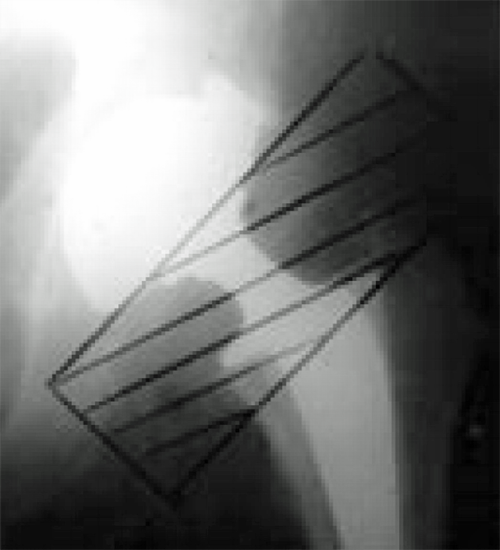
  • Prophylactic radiotherapy reduces HO after THA. A single dose of 700 cGy can be given either 4 hours preoperatively or within 72 hours (Figure 5). Both preoperative radiation and postoperative radiation were found to be equally effective at the hip, and no specific differences in complications were noted.
  • The mechanism of action is thought to be due to inhibition of the fast-dividing osteoprogenitor cells that are present within the first week after surgery.
  • These then differentiate into mature cell types such as osteoblasts. The lower mitotic rate of mature cells protects them from the damage to DNA caused by radiation.
  • Radiation exposure is limited to the soft tissues immediately around the hip joint, and ingrowth surfaces must be appropriately shielded.
  • Although no cases of malignancy after prophylatic radiation have been reported, this is a theoretical risk to consider.
  • Additional side effects include progressive soft tissue contracture, delayed wound healing, non-union and inhibited ingrowth of press fit implants.
BS3HO 5.png

Figure 5. The radiation field after THA.

NSAIDs versus radiotherapy

  • Pakos and Ioannidis (Int J Radiat Oncol Biol Phys 2004; 60: 888–895) performed a meta-analysis and suggested radiotherapy was more efficient than NSAIDs at preventing Brooker grade 3–4 HO.
  • The overall difference in absolute risk was, however, small.
  • Other studies have reported no difference between NSAIDs and radiotherapy in preventing heterotopic ossification.
  • The decision to use radiotherapy or NSAIDs medication for prophylaxis can be tailored to individual patient conditions.

Combination therapy

  • Radiotherapy and NSAIDs in combination may offer a potential advantage and three retrospective reviews have suggested lower incidence compared to single therapy, but further evidence is required.
Previous
Next

QUESTION 1 OF 3

Risk factors for heterotopic ossification around the hip include

QUESTION ID: 1040

1. Ankylosing spondylitis
2. Cemented arthroplasty
3. Female gender
4. Posterior approach
5. Rheumatoid arthritis

QUESTION 2 OF 3

Risk factors for heterotopic ossification include

QUESTION ID: 1041

1. Anaesthesia
2. Blood transfusion
3. Blood type
4. BMI
5. Prosthetic design

QUESTION 3 OF 3

Concerning HO and hip resurfacing.

All the following are TRUE EXCEPT

QUESTION ID: 1042

1. A sterile plastic drape can decrease the incidence of HO
2. Bone debris from femoral head reamings can stimulate the formation of HO
3. Lower rates of heterotopic ossification have been associated with hip resurfacing as compared to THA
4. The osteogenic precursor cells are derived from local tissue rather than blood-borne cell lines
5. The posterior approach is associated with the lowest incidence of HO

Further Reading

  • 1. Board TN, Karva A, Board RE, Gambhir AK, Porter ML. The prophylaxis and treatment of heterotopic ossification following lower limb arthroplasty. J Bone Joint Surg Br 2007; 89-B(4): 434–440.
  • 2. Kaplan FS, Glaser DL, Hebela N, Shore EM. Heterotopic ossification. J Am Acad Orthop Surg 2004; 12: 116–125.
  • 3. Ranganathan K, Loder S, Agarwal S, et al. Heterotopic ossification: basic-science principles and clinical correlates. J Bone Joint Surg Am 2015; 97(13): 1101–1111.
  • 4. Pape HC, Marsh S, Morley JR, Giannoudis PV. Current concepts in the development of heterotopic ossification. J Bone Joint Surg [Br] 2004; 86-B: 783–787.

References

  • 1. Mellema, J., et al., Pulsed Lavage in Cementless Total Hip Arthroplasty Reduces the Incidence of Brooker Grade 3 and 4 Heterotopic Ossifications. Open Journal of Orthopedics, 2011. 1(2): p. 9-13.
  • 2. Eggli, S. and A. Woo, Risk factors for heterotopic ossification in total hip arthroplasty. Archives of orthopaedic and trauma surgery, 2001. 121(9): p. 531-535.
  • 3. Shields, J.S., et al., Does a plastic drape reduce incidence of heterotopic ossification after hip resurfacing? Clinical Orthopaedics and Related Research¬Æ, 2011. 469(6): p. 1606-1613.
  • 4. Cohn, R.M., R. Schwarzkopf, and F. Jaffe, Heterotopic ossification after total hip arthroplasty. American journal of orthopedics (Belle Mead, NJ), 2011. 40(11): p. E232.
  • 5. DeLee, J., A. Ferrari, and J. Charnley, Ectopic bone formation following low friction arthroplasty of the hip. Clinical orthopaedics and related research, 1976. 121: p. 53.
  • 6. Maloney, W., et al., Incidence of heterotopic ossification after total hip replacement: effect of the type of fixation of the femoral component. The Journal of bone and joint surgery. American volume, 1991. 73(2): p. 191.
  • 7. Iorio, R. and W.L. Healy, Heterotopic ossification after hip and knee arthroplasty: risk factors, prevention, and treatment. Journal of the American Academy of Orthopaedic Surgeons, 2002. 10(6): p. 409-416.