TOPIC DETAILS

  Team Member Role(s) Profile
Paul Banaszkiewicz Paul Banaszkiewicz Section Editor
James Donaldson James Donaldson Segment Author
  • Infection is a condition in which pathogenic organisms multiply and spread within body tissues. This usually gives rise to the classical signs of:
  • redness
  • swelling
  • heat
  • pain
  • and loss of function
  • Osteomyelitis causes bone death, soft tissue compromise, functional impairment, systemic illness and considerable morbidity.
  • Osteomyelitis is defined as inflammation of the bone caused by an infecting organism. It is nearly always seen in children (with the exception of immunocompromised adults).

Aetiology

  • Infection generally arises from three sources:
  1. Haematogenous spread of bacteria is most common.
  • Bacteraemia results in the deposition of bacteria usually in the metaphysis of bone (most commonly around the proximal tibia or distal femur in children and vertebra in adults).
  • More common in those older than 60 years or younger than 20 years.
  • A single pathogen is usually isolated with the most common organism being Staphylococcus aureus.
  1. Contiguous spread caused by surgery, trauma or implant.
  2. Secondary to vascular insufficiency, usually from a soft tissue infection in patients with diabetes mellitus:
  • Usually caused by multiple organisms.
  • Local factors and the host response (extremes of age, malnutrition, immunosuppression, diabetes) will contribute to the subsequent signs and symptoms.
  • The causal organism can be identified by blood cultures in approximately 50% of patients. Staphylococcus aureusremains the commonest causative organism but others are prevalent and vary depending on age.

Organisms

  • Infants <1 year:
  • Group B Strep
  • S. aureus
  • Escherichia coli
  • Children 1–16 years:
  • S. aureus
  • Group A Strep
  • Haemophilus influenza
  • Adults:
  • S. aureus
  • Staphylococcus epidermidis
  • Gram –ve bacilli
  • Special cases:
  • Salmonella in sickle cell disease
  • Pseudomonas in IVDUs

Pathology (Figure 1)

  • The predilection for the metaphysis is thought to be due to the relative vascular stasis from hairpin loops in the nutrient artery, which favour bacterial colonisation. 
  • In neonates there is still a connection between the epiphysis and the metaphysis, and infection can settle in the end of the bone. 
  • Joint infection (septic arthritis) and growth disturbance can result. In older children the growth plate acts as a barrier to the spread of infection and acute osteomyelitis is usually confined to the metaphysis.

BS3INFECTIONBONESOFTTISSUE 1.png

Figure 1. Hairpin loops nutrient artery
  • There is a characteristic pattern of:
  • Inflammation.
  • Suppuration with pus formation by day 2 to 3.
  • A rise in intra-osseous pressure causes tissue necrosis and further vascular stasis. Oedema and thrombosis of local blood vessels may result in local bone lysis and necrosis allowing the infection to spread within the metaphysis.
  • Dead bone may separate as “sequestra.”
  • Reactive new bone forms at the end of the second week which slowly thickens forming an “involucrum” which encloses the infected tissue and sequestra.
  • Resolution and healing is the final stage if the infection is appropriately treated. 
  • Alternatively chronic infection may persist.

Clinical features

  • Pain with localised tenderness as well as fever and malaise are commonplace. The child may be refusing to use that limb and there may be a recent history of infection elsewhere. Local swelling and redness are later signs as the soft tissues become involved. In neonates the constitutional disturbance may be mild (failure to thrive and irritability).

Investigations

  • Blood tests: C-reactive protein (CRP) and erythrocyte sedimentation rate (ESR) are usually raised.
  • X-rays are often normal initially.
  • Ultrasound may show a subperiosteal collection.
  • Bone scan is sensitive but not specific.
  • Magnetic resonance imaging (MRI) is extremely sensitive and will show periosteal reaction, soft tissue involvement, and bone marrow changes.

Radiographs

  • In the early phase of acute osteomyelitis, i.e. the ?rst 2–3 days, plain radiographs are usually normal but over 6–7 days successively localised osteopenia, bone destruction, cortical breaches, periosteal reaction and a developing involucrum will become apparent.
  • Visible sequestra may appear at around 10 days.
  • Over several weeks, the whole bone will become generally osteopenic due to disuse. Any area of cortical bone, which remains without osteopenia, is likely to be avascular.

Differential diagnosis

  • Cellulitis
  • Septic arthritis
  • Acute rheumatic arthritis/Still’s disease
  • Sickle cell crisis
  • Necrotising myositis or fasciitis
  • Gaucher’s disease
  • Bone or soft tissue tumour

Treatment

  • Multidisciplinary approach.
  • Supportive treatment includes analgesia, intravenous fluids and splintage of the limb.
  • Antibiotics:
  • Commencement of broad-spectrum intravenous antibiotics with recognised anti-staphylococcal and streptococcus activity immediately after culture specimens (blood cultures or bone biopsy). 
  • Once an organism is identified the antibiotics can be modified accordingly.
  • Antibiotics are usually required for at least 6 weeks.
  • The earlier the infection is treated, the higher the chance of cure.
  • Surgery may be required when:
  • Significant sub-periosteal or soft tissue collections are present and these need to be drained.
  • To debride all dead bone and ischaemic tissues.
  • To treat recurrent disease or disease that is not responding to antibiotic treatment.

Principles of surgery

  • Early aggressive management along with optimisation of the host medical and nutritional status, with a focus on wide margins excision and debridement of all affected tissues.
  • When debriding healthy bleeding tissue should be visualised at all boundaries of the surgical site, followed by thorough irrigation.
  • Stage I infections limited to the medullary canal may be treated with intramedullary reaming.
  • Stage II infections can be addressed with soft tissue debridement and decortication of the bone adjacent to the infection.
  • Stage III osteomyelitis usually requires debridement of soft tissue, sequestrum and cortex, as well as decompression of the involved medullary canal.
  • Stage IV infections are the most challenging to treat surgically because they most often require multiple staged procedures and necessitate osseous stabilisation. 

Complications

  • Septicaemia
  • Metastatic infection
  • Septic arthritis
  • Recurrent infection – depends on the site and time to treatment
  • 20% recurrence around the knee
  • 50% in metatarsal
  • Early diagnosis 92% cured; late diagnosis 25% cured
  • Altered bone growth, deformity and limb length discrepancy
  • Pathological fracture
  • Chronic osteomyelitis
  • This is more insidious with fewer symptoms. Pain may be present for some weeks with minimal symptoms. The relative mildness is thought to be due to a less virulent organism or higher innate resistance of the patient.
  • Blood tests may be normal in 50% of cases. The classical radiograph may show a cavity, surrounded by a halo of sclerosis (the Brodie’s abscess). The most common sites are again the proximal tibia or distal femur. 
  • A benign or malignant bone tumour must be excluded and often a biopsy is needed. Microbiology is positive in 50% of cases and is almost always Staphylococcus aureus.
  • Treatment may be non-surgical with immobilisation and antibiotics. Surgery is indicated if the diagnosis is in doubt or if there is a lack of response radiographically or biochemically to prolonged antibiotic treatment.
  • Historically this often followed acute osteomyelitis. These days it is a more common sequel to an open fracture or surgical procedure.

Clinical features

  • Systemic features of infection may or may not be present. 
  • There may be localised pain and discharge.
  • Tissues may be thickened and indurated.
  • In long-standing cases joint contractures and bone deformity may occur.
  • The specific symptoms will depend on the physiological condition of the host, the site of involvement, the functional impairment and the extent of bone necrosis. 

Imaging

  • Radiographs may show: 
  • bone resorption 
  • periosteal thickening
  • osteoporosis
  • sequestra (Figure 2) and the surrounding involucrum

BS3INFECTIONBONESOFTTISSUE 2.jpg

Figure 2. Sequestrum of chronic osteomyelitis in tibia
  • Bone scan, MRI, computed tomography (CT) and sonograms may all be helpful.
  • Although several classification systems exist the two most widely used systems are those of Waldvogel and Cierny–Mader.

Waldvogel classification

  • This system is based on time course and aetiology.
  • Time course is either acute or chronic.
  • Aetiology is either haematogenous or secondary to a contiguous focus of infection.
  • The final part of the classification is the presence or absence of generalised vascular disease.
  • One strength of the classification is that it recognises the importance of poor tissue perfusion in the severity of osteomyelitis.
  • The main weaknesses are that it fails to assess the stage or severity of the treatment and does not directly guide treatment. It also ignores infection due to open trauma or surgery.

Cierny–Mader staging classification (Figure 3)

  • This system remains the most clinically relevant and stratifies hosts into three categories based on physiological comorbidities and designates four anatomical stages of infection that are combined to produce 12 classifications.
  • Stage 1: Medullary in which the infection is confined to the medullary canal of the bone.
  • Stage 2: Superficial in which the infection involves the cortical bone and may be caused by spread from a contiguous focus of infection or direct inoculation.
  • Stage 3: Localised in that the bone is stable and infection does not involve the whole circumference, although both cortical and cancellous bone may be involved.
  • Stage 4: Diffuse osteomyelitis that involves the entire thickness of the bone and impairs its stability.
  • With host type A is normal, type B is impaired by one or more systemic (drug use, age, diabetes mellitus, malignancy and malnutrition) or local disease process (chronic oedema, fibrosis from radiation, scarring) and type C describes a host so severely compromised that the risk of radical treatment outweighs its likely benefit.

BS3INFECTIONBONESOFTTISSUE 3.png

Figure 3. Anatomical classification of adult osteomyelitis

Treatment

  • Chronic osteomyelitis is difficult to treat and antibiotics alone are not sufficient. A variety of surgical techniques may be needed and a specialist bone infection centre should be involved.
  • Surgical debridement:
  • Dead bone, infected soft tissue, purulent material, sinus tracks and foreign material or metalwork all need to be removed.
  • Bone loss will need reconstruction:
  • Bone graft or limb reconstruction with an external fixator with larger defects.
  • Soft tissue:
  • May require local tissue flaps or free flaps.
  • Antibiotics:
  • Bone cultures and microbiology samples should be taken prior to any antibiotic treatment.
  • Local administration of antibiotic in cement, implants or via an irrigation system may be helpful.
  • Tuberculosis is most common in developing countries with crowding, poor sanitation and malnutrition. In the developed world its incidence is on the increase due to an increasingly elderly population, global travel and immunosuppressive disease states (especially HIV). Only a small number of patients (3–5%) with tuberculosis will have osteoarticular involvement, and half of these will have spinal disease.

Pathology

  • Mycobacterium tuberculosis is an aerobic bacterium that enters the body via the lungs or gastrointestinal tract. It causes a granulomatous reaction, which is associated with tissue necrosis and caseation. The primary focus of infection is usually in the lung and regional lymph nodes. Musculoskeletal involvement is via haematogenous spread.
  • Musculoskeletal disease may begin as synovitis or osteomyelitis. At this stage soft tissue swelling is seen with a reduction in movement. Degenerate changes and eventual joint destruction ensue. Healing is often by fibrous ankylosis.

Clinical features

  • Constitutional:
  • Low grade fever
  • Night sweats
  • Weight loss
  • Anorexia
  • Malaise
  • Musculoskeletal:
  • Swelling and stiffness
  • Restricted movement and pain
  • Deformity
  • Sinuses
  • Neurological deficit (spine)
  • Investigations
  • X-ray:
  • Soft tissue swelling
  • Peri-articular osteopenia, erosions
  • Little or no periosteal reaction
  • Spine: bone erosion and collapse around a narrowed or destroyed disc space
  • Bloods: raised ESR
  • Positive Mantoux test
  • Acid-fast bacillus (AFB) in synovial fluid
  • Tissue biopsy is the most sensitive for AFB

Treatment

  • Chemotherapy:
  • Specialist anti-tuberculosis treatment is essential usually with multiple therapies to limit resistance.
  • Surgery is seldom necessary, except:
  • Emergency spinal decompression and stabilisation in cases of impending or actual paraplegia.
  • Joint arthrodesis or arthroplasty once the disease is quiescent. Anti-tuberculous treatment should be given pre and postoperatively when joint replacement surgery is performed.
  • Deep subcutaneous involvement
  • Often indistinct borders
  • Group A Streptococcus
  • S. aureusoccasionally
  • Treat with antibiotics

BS3INFECTIONBONESOFTTISSUE 4.jpg

Figure 4. Cellulitis affecting hand
  • Superficial, well demarcated
  • Red, raised painful plaque
  • Group A Streptococcus
  • S. aureus
  • Treat with antibiotics

BS3INFECTIONBONESOFTTISSUE 5.jpg

Figure 5. Erysipelas
  • Involves the muscle fascia 
  • Aggressive
  • Group A Strep
  • Extensive surgical debridement

BS3INFECTIONBONESOFTTISSUE 6.jpg

Figure 6. Before surgical debridement

BS3INFECTIONBONESOFTTISSUE 7.jpg

Figure 7. After surgical debridement
 
  • Affects muscle
  • Pain, oedema and discharge
  • Clostridium perfringens or specticum
  • Debridement and fasciotomy with or without hyperbaric oxygen
  • Postoperative wound discharge and erythema (Figures 8 and 9)
  • Species vary
  • S. aureus
  • S. epidermidis
  • MRSA
  • Group A Strep
  • Treated with antibiotics with or without wound debridement

BS3INFECTIONBONESOFTTISSUE 8.jpg

BS3INFECTIONBONESOFTTISSUE 9.jpg

Figure 8 and Figure 9. Post op wound infection

  • Swollen and tender, sausage shaped finger (Figure 10)
  • Flexed position and pain with passive movement
  • S. aureus
  • Splintage and antibiotics if caught very early. Usually surgical drainage is required

BS3INFECTIONBONESOFTTISSUE 10.jpg

Figure 10. Suppurative tenosynovitis.Pyogenic flexor tenosynovitis of the index finger of the right hand.

  • Infected paronychial fold along a nail
  • S. aureus
  • Drainage of pus with or without antibiotics

BS3INFECTIONBONESOFTTISSUE 11.jpg

Figure 11. Paronychia

  • Human (Figure 12) or animal
  • Multiple infective species
  • Broad spectrum antibiotics. Often wound washout is needed for hand bites (“fight bites”)
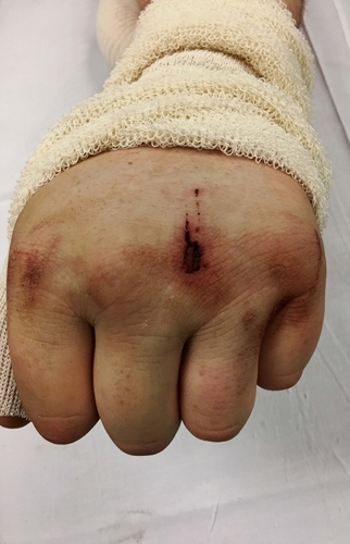
BS3INFECTIONBONESOFTTISSUE 12.jpg

Figure 12. Clenched-fist injury. Also known as a fight-bite injury, this serious injury typically is characterized by a 3- to 5-mm laceration on the dorsum of the hand or overlying the metacarpophalangeal joint.

  • Neuroparalytic disease from contaminated wounds
  • Clostridium tetani
  • Tetanus immunoglobulin if vaccination status unknown
  • Polio is an acute viral disease that affects the anterior horn cells of the spinal cord and brain stem, resulting in asymmetrical lower motor neuron palsy. It is a rare disease in developed countries following widespread vaccination.

Clinical features

  • Only a small proportion of patients exhibit any symptoms but several recognised stages are seen:
  • Acute illness: fever, headache, flexed joints with pain when they are stretched.
  • Flaccid paralysis: peaks at 2–3 days, may give rise to difficulty in breathing.
  • Recovery: from 10 days to up to 2 years.
  • Residual paralysis: asymmetric weakness may lead to joint deformities.
  • Post-polio syndrome: progressive muscle weakness in both old and new muscle groups due to “neural fatigue.”

Treatment

  • Physiotherapy and surgical appliances is the mainstay of treatment.
  • Surgery, when necessary, is aimed at restoring function – tendon transfers, osteotomies, joint stabilisations.
  • Infection limited to the intervertebral disc is rare. In adults it is usually a result of an intervention or surgery. In children it is from haematogenous spread. The vertebral end-plates are rapidly destroyed and the infection can progress into the vertebral body. Acute back pain, muscle spasm, limitation of movements and systemic infective features may be seen.
  • Treatment is with prolonged antibiotics. Surgery is seldom indicated except when there is cord compression from abscess formation.
  • Is defined as inflammation of the synovial membrane with purulent effusion into the joint capsule and is caused by bacteria within the joint space. The use of antibiotics has virtually eliminated mortality but permanent joint damage may result if it is not treated within 24–48 hours. There are approximately 20,000 cases per year in the UK.

Aetiology

  • Sources of infection include:
  • Direct inoculation from a penetrating wound, surgery or aspiration.
  • Spread from adjacent infection or abscess.
  • Haematogenous spread from a distant site.
  • Predisposing factors include rheumatoid arthritis, immunocompromise, advanced age, chronic debilitating conditions, intravenous drug abuse.

Pathology

  • The normal joint does have protective components with some phagocytic and bactericidal activity. However, a synovial joint is highly vascular and has no basement membrane to act as a barrier to microbes. In addition, previously damaged joints may demonstrate neovascularisation and have increased adhesion factors making them more susceptible to infection.
  • The organism crosses the synovial membrane and the acute inflammatory cascade is initialised. As pus appears in the joint, destructive enzymes within the exudate begin to erode the articular cartilage. In the early stages this process is reversible. Within the next 24–48 hours marked synovial proliferation, infiltration by mononuclear cells and granulation tissue develop and permanent damage may ensue.

Clinical features

  • The classic presentation is of an acutely hot and swollen joint with systemic signs of infection. It may differ according to the age of the patient. In the newborn the emphasis is on septicaemia rather than joint pain; in children and adults the typical features often include:
  • Severe pain, worse with any degree of movement.
  • Fever, malaise and systemic signs of sepsis.
  • A red, hot and swollen joint with an effusion.
  • In the immunocompromised a high index of suspicion is needed, as there may be few systemic symptoms and signs.

Investigations

  • Raised white cell count, CRP and ESR. 
  • Blood cultures may be positive.
  • X-rays may help in excluding other causes (Figure 13).

BS3INFECTIONBONESOFTTISSUE 13.jpg


  • Ultrasound is useful and an aspiration can be performed at the same time, especially for deeper joints like the hip.
  • MRI (Figure 14).

BS3INFECTIONBONESOFTTISSUE 14.jpg

Figure 14. Septic arthritis of the right hip

  • Synovial fluid examination is the gold standard:
  • A white cell count greater than 50,000 per ml is usually suggestive of infection.
  • The Kocher criteria for a child with a painful hip include:
  • Raised CRP
  • Raised white cell count 
  • Inability to bear weight
  • Pyrexia
  • Four of the criteria are 99% sensitive for septic arthritis; three are 93% sensitive; two are 40% sensitive and one is 3% sensitive.

Differential diagnosis

  • Acute osteomyelitis. The two may co-exist in young children – the treatment is the same.
  • Trauma: synovitis or haemarthrosis.
  • Irritable joint (the child will be systemically well).
  • Crystal monoarthropathy.
  • Haemophiliac bleed.

Management

  • Treatment may be started as soon as an aspirate is taken. The principles are similar to acute osteomyelitis:
  • Supportive treatment with IV fluids, analgesia and splintage.
  • Surgical drainage and washout is standard in the UK. This is usually an open procedure. A further washout may be needed if symptoms do not settle within 24–48 hours.
  • Antibiotics: initially best guess broad spectrum intravenous antibiotics are started and changed when microbiological sensitivities are known. Intravenous antibiotics may be converted to oral antibiotics once signs of sepsis have resolved and then continued for 6 weeks. The timescale is controversial and may be shorter or longer depending on the response.

Prognosis

  • 30% of adults with septic arthritis are left with reduced range of movement or chronic pain post-infection.
  • Bone destruction and dislocation (especially the hip) may occur.
  • Cartilage destruction may lead to ankylosis or secondary osteoarthritis. 
  • Growth disturbance and deformity occur in children.
  • Predictors of poor outcome include:
  • Age >60 years.
  • Infection of hip or shoulder joint.
  • Underlying rheumatoid arthritis.
  • Positive findings on synovial fluid cultures 7 days after starting antibiotics.
  • Delay in treatment.
Previous
Next

QUESTION 1 OF 2

Which of the following is not part of Kocher’s criteria for differentiating septic arthritis of the hip?

QUESTION ID: 1292

1. CRP
2. Erythrocyte sedimentation rate
3. Fever
4. Non-weight bearing
5. White cell count

QUESTION 2 OF 2

A 60-year old patient with back pain has an MRI scan suggesting tuberculous spondylodiscitis..Routine microbiology culture tests are negative.There is no concern about malignancy.
Which of the following is correct?

QUESTION ID: 1293

1. Mycobacterium TB appears as bright red colonies on Lowenstein–Jensen culture medium
2. Obligate anaerobic, acid-fast bacilli would be seen in TB
3. TB of the spine is the least common form of musculoskeletal TB
4. The presence of skip lesions on the MRI suggests this is not TB
5. Venous impregnation is the most likely route of spread