TOPIC DETAILS

  Team Member Role(s) Profile
Paul Banaszkiewicz Paul Banaszkiewicz Section Editor, Segment Author
Chris Ghazala Christopher George Ghazala Segment Author

Section editor: Paul Banaszkiewicz

Segment author: Christopher George Ghazala; Paul Banaszkiewicz

Document history:6/07/2019

  • Ligament injuries are common and management requires an understanding of their mechanical properties and healing mechanisms.
  • Ligaments are short flexible dense bands of collagenous tissues (fibres) that span a joint and then become anchored to the bone at either end.
  • Prevent excessive joint motion.
  • Connect bone to bone.
  • Passively stabilise joints.
  • Offer early and increasing resistance to tensile loading over a narrow range of joint motion.
  • Contribute to proprioception and position sense.
  • The fibres in ligaments are arranged in a wavy pattern (crimp) increasing their capacity to absorb tension.
  • This arrangement contrasts with tendons in which the arrangement of collagen fibres is essentially parallel.
  • A less parallel arrangement of collagen fibres in ligaments allows them to resist tensile stresses not only in one specific direction but also smaller stresses in other directions.
  •  Ligaments have the same general composition as tendons but with a few key differences.
  • Due to the organisational pattern of fibres, not all fibres are stretched when loaded along the main fibre axis.
  • Ligaments are less strong than tendons.
  • Similar to tenocytes, ligaments have fibroblasts that are found within the ligamentous substance aligned with the collagen fibrils.

Table 1. Comparison of tendons and ligaments


Ligaments

Tendons

% of collagen

Lower

Higher

% of ground substance

Higher

Lower

Organisation

More random

Organised

Metabolic activity

Higher

Lower

Orientation

Weaving pattern

Long axis direction


Table 2. Comparison of composition between tendon and ligament

Component

Ligament

Tendon

Cellular material


Fibroblasts

20%

20%

Extracellular material (matrix)


Water

60–80%

60–80%

Solids

20–40%

20–40%

Collagen

70–80%

Slightly higher

Type 1

90%

95–99%

Type 3

10%

1–5%

Ground substance

20–30%

Slightly less

Elastin

Up to 2x collagen

Scarce

   
  • Both ligaments and tendon display stress-strain behaviour that is time and rate dependent. The three main features of viscoelastic properties are:
  • Hysteresis: as the loading rate increases, the stiffness of ligaments and tendons increases, storing more energy to failure and undergoing more elongation to failure.
  • Stress relaxation: if a tendon or ligament is held at a constant length (strain) by a load (stress), the stress required to maintain that strain reduces. This reduces rapidly during the first 6–8 hours of loading and then more slowly over the next few months.
  • Creep: if a tendon is held by a constant load (stress), with time the length (strain) in the tendon will increase. This strain increases quickly at first but then increases more slowly. This phenomenon is taken advantage of by the application of plaster casts or braces to correct deformity, e.g. in scoliosis or clubfoot.
  • This is demonstrated when the loading of a sample is stopped below the linear region of the load–deformation curve and the amount of load remains constant over an extended period of time (i.e. the amount of elongation is constant). The deformation increases rapidly at first and then gradually more slowly.
  • Occurs when a viscoelastic material that is subjected to a constant load continues to stretch over time.
  • It is a time-dependent elongation of a tissue when subjected to a constant stress.

BS2LIG 1.jpg

Figure 1. Creep

  • This is demonstrated when the loading of a sample is stopped below the linear region of the load–deformation curve and the sample is maintained at a constant length over an extended period of time (i.e. the amount of elongation is constant). The load decreases rapidly at first and then gradually more slowly.
  • Stress relaxation is a time-dependent, viscoelastic process similar to creep, but it refers to the decay of applied stress under conditions of constant strain (in contrast to creep, which is a time-dependent strain under conditions of constant stress).
  • A reduction in the internal resistance (stress) over time when a viscoelastic material is stretched and fixed in length without further deformation.

BS2LIG 2.jpg

Figure 2. Stress relaxation

  • Ligaments are covered by a vascular and cellular overlying layer called the epiligament, which is often indistinguishable from the actual ligament.
  • The epiligament contains sensory and proprioceptive nerves, the larger percentage of which is located closer to the bone ligament insertion sites.
  • Force-elongation curves involve mounting a specimen in a machine whereas stress/strain curves have been normalised with respect to specimen dimensions.
  • Force–elongation curves are essentially the same as stress/strain curves.
  • Region 1: 'primary' or 'toe' region. Elongation occurring here is believed to be the result of straightening in the wavy pattern of relaxed collagen fibres. A small increase in load/stress causes a relatively large change in length/strain. The stiffness/slope of the graph/modulus of elasticity increases during this part of the curve.
  • Region 2: 'secondary' or 'linear' region. The fibres straighten out and the stiffness of the specimen has increased and become constant. Hence the slope of the graph is constant. = elastic region.
  • Region 3: the end of the linear region there is early sequential failure of a few very stretched collagen fibres, causing dips. The load/stress at which this occurs is called the yield point.
  • Point 4: the ultimate stress/strength is the maximum load/stress that can be achieved before the ligament/tendon completely fails.A series of small drops as ultimate failure occurs, due to sequential failures of fibre bundles.

BS2LIG 3.png

Figure 3. Force elongation curve of ligament 

  • There are several variants of the stress/strain graph of both  ligaments and tendons.
  • Often the stress/strain graph is labelled as a composite tendon/ligament
  • Choose a straightforward reproducible diagram easy to draw and explain.
  • The stress/strain graph of both ligament and tendon are similar as both have the same sort of microscopic crimp, only this is greater in ligaments. As a result there is a more non-linear load–deformation curve earlier on.

Ligaments have lower mechanical properties-the ultimate tensile strength and elastic modulus are less than tendons.

BSLIG 4.jpg

  • When a ligament is subjected to loading that exceeds the physiological range (injury due to high levels of stress), either microfracture occurs even before the yield point is reached (i.e. partial rupture of a ligament) or if the yield point is exceeded, the ligament will undergo gross failure (complete rupture).
  • Ligaments are classified in three grades of injury severity:
  1. First degree: minimal symptoms, some pain is felt but no joint instability is noted. Some micro failure of collagen fibres but no macroscopic disruption.
  2. Second degree: severe pain and some joint instability. Partial ligament rupture.
  3. Third degree: severe pain during the event of injury but less pain afterwards. The joint is completely unstable.
  • Collagen synthesis and degradation proceed simultaneously, as in other types of wound healing, but collagen content increases.
  • Initial collagen is type 3, although later composition changes to predominantly type 1 collagen.
  • Glycosaminoglycans also increase in the early phase of wound healing.
  • With remodelling and maturation, contents gradually return to normal.
  • Injured ligaments sometimes demonstrate the ability to contract or to “tighten up.” (Rat medial collateral ligaments Z-lengthened are able to contract to normal tightness in three weeks.)
  • There is an associated increase in the amount of actin as measured by immunofluorescent staining (actin is same contractile protein found in thin filaments of muscle sarcomeres and in the cytoplasm of mobile cells).
Previous
Next

QUESTION 1 OF 7

A 22-year-old male has undergone ACL reconstruction and has been referred on to physiotherapy.
When comparing open versus closed chain post op exercises

QUESTION ID: 1050

1. Open chain exercises are safer than closed chain exercises because they place less strain on the ACL graft.
2. Open chain exercises are the preferred choice rather than closed chain exercises in ACL rehabilitation
3. Open chain exercises have better knee extensor strength
4. Open chain exercises have better pain scores
5. Open chain exercises have improved active knee flexion

QUESTION 2 OF 7

Compared to tendons ligaments have:

QUESTION ID: 1051

1. Greater tensile strength
2. Higher content of collagen
3. Lower elastic modulus
4. Lower proteoglycan content
5. Lower water content

QUESTION 3 OF 7

A ligament is loaded and unloaded several times with the load /elongation curve being shown in figure 1.1

Regarding figure 1.1:

SBALigament 1.png

QUESTION ID: 1053

1. After about 20 cycles the loading and unloading curves do not change anymore but they are still
2. Decrease in stress becomes less with repeated loading
3. During cyclic loading and unloading the stress/strain curve shifts to the left
4. The amount of hysteresis under cyclic loading is increasing
5. The figure demonstrates pseudo-elasticity

QUESTION 4 OF 7

86.A 22-year-old male has undergone ACL reconstruction and has been referred on to physiotherapy.
When comparing open versus closed chain post op exercises

QUESTION ID: 2242

1. Open chain exercises are safer than closed chain exercises because they place less strain on the ACL graft.
2. Open chain exercises are the preferred choice rather than closed chain exercises in ACL rehabilitation
3. Open chain exercises have better knee extensor strength
4. Open chain exercises have better pain scores
5. Open chain exercises have improved active knee flexion

QUESTION 5 OF 7

88.Compared to tendons ligaments have

QUESTION ID: 2244

1. Greater tensile strength
2. Higher content of collagen
3. Higher metabolic activity
4. Lower proteoglycan content
5. Lower water content

QUESTION 6 OF 7

69. During ACL reconstruction surgery when a surgeon is preparing the ACL graft the biomechanics of ACL graft preconditioning are discussed
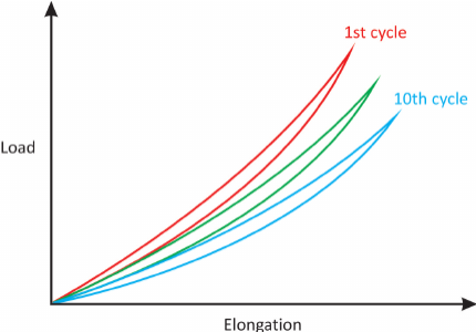
During cyclic loading and unloading of an ACL ligament(Figure 1)
cONDITIONING .png

Figure 1 During cyclic loading and unloading, the stress/strain curve shifts to the right. After 10 repetitions, the curve becomes reproducible. The amount of hysteresis under cyclic loading is reduced.


QUESTION ID: 2246

1. Displays greater stiffness at the 10th cycle
2. Leads to the ligament resembles an elastic spring
3. The amount of hysteresis is increased
4. The diagram demonstrates  pseudo-plasticity which represents the nonlinearity of ligament stress strain behaviour
5. The stress/strain curve shifts to the left

QUESTION 7 OF 7

60.Which of the following statements is true of the differences between a ligament and a tendon?

QUESTION ID: 3257

1. Ligaments have more highly aligned collagen fibres than tendons
2. Tendons are more cellular than ligaments
3. Tendons are more metabolically active than ligaments.
4. Tendons contain more elastin than ligaments.
5. Tendons display less hysteresis than ligaments

Further Reading

  • 1. Fralinger DJ, Kaplan DJ, Weinberg ME, Strauss EJ, Jazrawi M. Biological treatments for tendon and ligament abnormalities. JBJS Reviews 2016; 4(6): e5.