TOPIC DETAILS

  Team Member Role(s) Profile
Paul Banaszkiewicz Paul Banaszkiewicz Section Editor, Segment Author
Chris Ghazala Christopher George Ghazala Segment Author
  • Three basic muscle classes: skeletal, cardiac and smooth muscle. Two basic muscle fibre types: fast-twitch and short-twitch.
  • Skeletal muscle is under voluntary control and mediates locomotion, respiration, phonation and posture.
  • Muscle consists of numerous multinucleated cells termed muscle fibres and each fibre is surrounded by an endomysium connective tissue layer. Groups of muscle fibres are called fascicles and these are encased by a further layer called the perimysium. Groups of fascicles form the main muscle substance and surrounding this muscle is an epimysium.
  • Muscle fibres are composed of numerous myofibrils, producing a striated histological appearance based on refraction under polarised light. The cell membrane of the muscle fibre is the sarcolemma and invaginations of this membrane towards the myofibrils give rise to T-tubules.
  • Each myofibril consists of regularly arranged thick and thin filaments.
  • Myofibrils are subdivided longitudinally into sarcomeres, limited by Z-lines at each end, thus Z-lines define the limits of each sarcomere; each sarcomere represents a contractile unit.
  • Striations are formed by the overlapping of thick and thin filaments, with the dark area of the A-band representing this area of overlap. Light bands, composed primarily of actin, lie on each side of the Z-line (I-bands) and extend from this region towards the centre of the sarcomere.

B2SMUSCLE 1.jpg

Figure 1. Elecronmicrograph of a myofibril.

  • The A-band lies between the two I-bands and contains thick filaments of primarily myosin (myosin-II) overlapping with thin actin filaments. In the centre of this A-band lies a light area of myosin, called the H-band; this is an area of relaxation with no overlap of thick and thin filaments.
  • Troponin-T binds tropomyosin and troponin-I inhibits the interaction with myosin and actin. When troponin-C binds Ca2+this causes disruption of the troponin-I and actin interaction, exposing the myosin-binding sites on actin.

BS2MUSCLE 2.jpg

Figure 2. Arrangement of the thick and thin filaments within a sarcomere during relaxation and contraction.  During contraction, the Z-lines of the sarcomere move closer together.

  • Myosin-II (~480 kDa) has two globular heads which form cross-bridges with actin via their actin binding site and myosin filaments are attached to the Z-lines by a titin cytoskeleton. It contains one heavy chain pair and two pairs of light chains, and the large chains form a large globular head which projects towards the actin filament.
  • Thin filaments are polymers formed by actin (G-actin or globular actin) into a two-stranded helix called filamentous actin; thin filaments also contain molecules of troponin and tropomyosin. Nebulin and tropomyosin run along the length of the filament. Tropomyosin runs between the two actin chains and covers the myosin-binding sites for the myosin-actin cross-bridge. Troponin consists of troponin-T, troponin-I and troponin-C, and regulates the tropomyosin molecules on actin and exposure of the myosin-binding sites.
  • Troponin-T binds tropomyosin and troponin-I inhibits the interaction with myosin and actin. When troponin-C binds Ca2+this causes disruption of the troponin-I and actin interaction, exposing the myosin-binding sites on actin.
  • Muscle contraction occurs when the action potential spreads across the motor end-plate of the muscle fibre and down the T-tubules of the sarcolemma, resulting in the release of Ca2+via the terminal cisternae of the sarcoplasmic reticulum, initiating contraction of the myofibrils. This process is known as excitation–contraction coupling.
  • Binding of the myosin head to actin results in the movement of the actin filaments towards the sarcomere’s centre and this process is dependent on adenosine triphosphate (ATP) confirmation changes at the myosin head. Overall, the sarcomere is shortened and the Z-lines are brought closer together, thus contracting the muscle fibre; contraction does not alter the overall length of the thick and thin filaments, just their overlap. The force generated by myosin is referred to as the cross-bridge cycle.
  • Muscle requires ATP for contraction, thus converting chemical into mechanical energy. ATP is derived from creatine phosphate, in which the enzyme creatine phosphokinase (CPK) catalyses the conversion of adenosine diphosphate (ADP) and creatine phosphate into ATP (and creatine). Other sources for ATP come from glycogen, fatty acids and triglycerides.
  • The sliding filament theory is the process of cross-bridge cycling in which myosin cross-bridging to actin results in pulling of the actin filament towards the centre of the sarcomere, thus shortening the myofibril and ultimately the muscle fibre.
  • Ca2+released from the sarcoplasmic reticulum binds to troponin-C (four Ca2+binding sites per molecule), resulting in exposure of the myosin-binding sites on actin.
  • Myosin binds with actin and undergoes conformational change, pulling actin towards the sarcomere centre (ratchet action). ATP is hydrolysed and inorganic phosphate (Pi) is released from myosin, causing release of the myosin head from actin.
  • ATP is partially hydrolysed to ADP and Pi by myosin, causing rococking of the myosin head, returning it to the resting state, completing the cycle. This cycle repeats until intracellular Ca2+is depleted. Rigor mortis is the result of permanent actin–myosin complexes due to insufficient ATP.
  • Providing the muscle is re-stimulated before it relaxes, intracellular Ca2+increases, thus magnifying the force of contraction – tetany.
BS2MUSCLE 3.jpg

Figure 3. Sliding filament theory.  (A) At rest, the myosin heads cocked and not bound to actin; actin is covered by tropomyosin.  (B) Calcium binds to the troponin-tropomyosin complex, causing exposure of the myosin binding sites on actin and cross-bridging.  (C) Myosin heads rotate and move actin over myosin, shortening the fibre and forming the power stoke.  (D) ATP binds to the myosin head, causing myosin to detach and (E) ATP is hydrolysed into ADP and Pi causing recocking of the myosn head from actin.

CASE BASED DISCUSSIONS

CBD Muscle physiology

Question:“Here is a piece of paper can you draw me a sarcomere (muscle cell)?

  • Sarcomere is the basic unit of muscle.
  • Contains actin (thin) and myosin (thick filaments).
  • Appropriately labels parts of the diagram, including: A (anisotropic) and I (isotropic) bands, etc.
  • Actin is bound to the Z line (German for “band in between”).

Question:“Tell me about the sliding filament theory of muscle contraction

  • Motor neurone discharges at neuromuscular junction.
  • This causes depolarization, and action potential travels along the T tubules.
  • At rest tropomyosin covers actin binding sites on the myosin protein filament, preventing cross-bridging.
  • Cleavage of ATP causes a conformational change in myosin head, allowing cross-bridging between actin and myosin.
  • Myosin head flexes, pulls the actin along.
  • Further ATP detaches the head and cycle repeated.
  • Cross-bridging allows actin filament to slide relative to myosin, which shortens sarcomere.

Question:“A lot of ATP is required for muscle contraction. Where does the energy come from? For instance, what happens in the first 60 secs when I go for a run?”

  • ATP is needed for muscle contraction
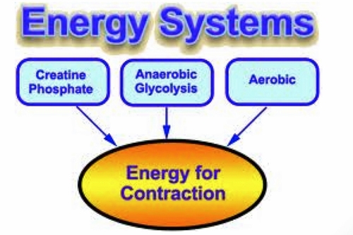
  • Three energy systems generate ATP
  1. ATP-CP system
  2. Anaerobic/Lactic acid system
  3. Aerobic system
  • ATP-CP system provides  an immediate pool of ATP available for a small number of muscle contractions.
  • Thereafter glycolytic pathway.
  • Finally an aerobic system via Krebs cycle.
 BS2MUSCLE 4.jpg

Figure 4. Energy systems 

Previous
Next

QUESTION 1 OF 4

Removed by super user

QUESTION ID: 1049

QUESTION 2 OF 4

68.The physiotherapist has left a message for you to contact them regarding the introduction of closed chain exercises for rotator cuff dysfunction.
Examples of closed chain exercises include

QUESTION ID: 2241

1. Bench press
2. Biceps curl
3. Latissimus pull down
4. Push ups
5. Triceps extensions

QUESTION 3 OF 4

87. Concerning type 1 and type 2 muscle fibres

QUESTION ID: 2243

1. Type 1 fibres are divided into types 1a and I b
2. Type 1 fibres have a high strength of contraction
3. Type I fibres tend to be smaller than type II in children and in adults who do not carry out strenuous activity although they increase in size with repeated physical exercise
4. Type II fibres are more prone to anatomic changes following altered energy demands than are type I fibres
5. Type II fibres have the smallest motor unit size

QUESTION 4 OF 4

53. When a sarcomere contracts

QUESTION ID: 3249

1. A band becomes shorter.
2. H zone stays the same.
3. I band stays at the same length.
4. M line shortens.
5. Z lines move closer together.

References

  • 1. Barrett K, Brooks H, Boitano S, Barman S. Ganog’s review of medical physiology, 24th edn. McGraw-Hill Medical, 2012.
  • 2. Berne RM, Levy MN, Koeppen BM, Stanton BA. Physiology, 5th edn. Mosby, St. Louis, USA, 2004.