TOPIC DETAILS

  Team Member Role(s) Profile
Paul Banaszkiewicz Paul Banaszkiewicz Section Editor
James Donaldson James Donaldson Segment Author
  • Is a disorder of incomplete spinal cord closure or secondary rupture of the developing cord due to hydrocephalus. 
  • Includes (see Figure 1):
  • Spina bifida occulta = defect in vertebral arch with confined cord and meninges
  • Meningocele = sac without the neural elements protruding
  • Myelomeningocele = protrusion of sac with neural elements
  • Rachischisis = neural elements exposed with no covering
  • Related to folate deficiency.
  • Incidence of laminar defects is 5%, cystic two to three/1000 live births.
  • Screened for in uterowith alphafeto protein and ultrasound scan (USS).
  • If one child affected risk increased by ten times.
  • Folic acid is routinely given to pregnant women.

BS3NMD 1.jpg

Figure 1. Spina Bifida

Spina bifida occulta

  • Midline defect in lamina, may have overlying dimple or hairy patch.
  • Occasional intraspinal anomalies, tethering of conus medullaris, diastomatomyelia, cysts/lipomas or cauda equine.
  • Function is related to the level of the defect and associated congenital abnormalities.
  • Commonest comorbidity is Arnold–Chiari malformation type II.
  • Sudden changes in function are associated with a tethered cord, hydrocephalus (70%) and hydromyelia.
  • Hydrocephalus:
  1. Distal tethering of cord may cause herniation of cerebellum through foramen magnum causing a cerebrospinal fluid (CSF) block.
  • Fractures are common (lower limb).
  • Latex allergy in 20–40% from multiple surgical procedures and ventricular–peritoneal (VP) shunt.

Genetics

  • Certain health problems in the mother, or medication used for their treatment, increase the susceptibility for a fetus to have a neural tube defect (NTD).
  • These include maternal diabetes, obesity, folate deficiency, or folate-antagonist anticonvulsants prescribed for epilepsy.
  • Most NTDs show multifactorial inheritance. However, they may also be seen as a feature in some chromosome or rare single gene disorders, usually in combination with other malformations.
  • A positive family history of NTD significantly increases the genetic recurrence risk (around 4% when one parent or a previous child has NTD).

Neurological dysfunction

  • Myelomeningocoele is always associated with neurological dysfunction below level.
  • Occult is usually associated with less severe neurological dysfunction.

Clinical features

Occulta 

  • Isolated laminar defects often in clinically normal people.
  • Hairy patch, dimple naevus, signifies more serious disease.
  • May present at any age with neuro symptoms, cauda equina or weakness.
  • X-rays show a laminar defect.
  • Bony midline ridge suggests diastomatomyelia.
  • Intra-spinal lesions shown on magnetic resonance imaging (MRI).

Cystica

  • Obvious defect at birth.
  • Abnormal baby posture in utero– muscle imbalance, hip dislocation, talipes, claw toes.
  • One third = complete lower motor neuron paralysis and loss of sensation and sphincter control below level.
  • One third = complete lesion at some level but distal segment of cord preserved – intact upper motor neuron lesions below.
  • One third = cord lesion incomplete so some movement and sensation preserved.
  • Muscle charting should be done within 24 hours to prevent worsening function due to traction of adherent nerves with growth.
  • L4 is the key level (quadriceps).
  • L1: Hip ER and flexed, equinovarus foot. Non-functional.
  • L2: Adducted and flexed hip, flexed knee, equinovarus foot. Non-functional.
  • L3: Adducted and flexed hip, recurvatum of the knee, equinovarus foot. Household ambulatory in knee–angle–foot orthosis (KAFO).
  • L4: Adducted and flexed hip, extended knee, cavovarus foot. Household plus ambulatory in ankle–foot orthosis (AFO).
  • L5: Flexed hip, limited flexion at the knee, calcaneal valgus. Community ambulatory in AFO.
  • S1: Foot deformities. Near normal mobility.

Treatment

Principles

  • Team approach.
  • Orthotics.
  • Surgery: balance muscles and deformities.

Early surgery

  • Skin closure to prevent drying and ulceration within 48 hours. 
  • Ventriculocaval shunt for hydrocephalus, to prevent brain damage.
  • Urinary problems: 90% of cases. Intravenous pyelogram (IVP) to detect urinary tract dilatation. Boys: penile appliance; girls: urinary diversion.
  • Passive stretching and strapping at 3 weeks for deformities.

Late principles

  • Stretching for first 6–12 months.
  • Open methods of correction of deformity.
  • Proximal deformity corrected before distal.
  • Never splints alone. Never for obtaining correction. Only for maintaining correction with stretching. 
  • Check for latex sensitivity prior to surgery/latex-free surgery.

Spine

  • Other abnormalities can occur and may need treatment, e.g. unsegmented bars, hemivertebrae, causing deformity. Kyphuscan be excised and fused. 
  • Deformities are often severe and progressive.
  • Bracing is not helpful.
  • Rapid progression can be associated with a tethered cord.
  • Severe and progressive deformities need.

Hip

  • Aim for hips straight enough to stand in calipers, flexible enough to sit.
  • If L1, all muscle groups paralysed therefore no problem.
  • If S1, pure flexion deformity treat with psoas lengthening.
  • If between, risk of dislocation is 50% by 2 years.
  • Treat with closed reduction and adductor tenotomy, psoas transfer from lesser to greater trochanter. 
  • Older children: varus osteotomy/inominate osteotomy/leave alone. 

Knee

  • Aim for a straight knee.
  • Quads weakness treated with a KAFO.
  • Flexion contractures treated with hamstring and posterior capsular release.
  • Recurvatum is rarely a problem. Treat with serial casting, quads lengthening and KAFO.

Foot and ankle

  • Aim for a plantigrade foot.
  • A floppy foot with complete paralysis just needs bracing and good footwear.
  • Deformities: tendon release if tendon paralysed and contracted, tendon transfer if tendon active but unopposed. 
  • Valgus foot is common. Treated with AFO, clubfoot release, posterior tibialis, Achilles and tibialis anterior lengthening.
  • Rigid clubfoot is common at L4. Treat with subtalar release, lengthening of tibialis posterior and Achilles and transfer of tibialis anterior to dorsal midfoot.
  • Vertical talus Rx.
  • Claw toes: flexor extensor transfer.

Prognosis

  • One third achieve independent walking, two thirds are wheelchair bound. 
  • Deteriorating neurological function due to:
  • Progressive hydrocephalus from shunt malfunction 
  • Hydromyelia from shunt malfunction 
  • Spinal cord tethering with growth 
  • Arnold–Chiari malformation 
  • HSMN 1, 2 and 3 are the most common.
  • Diagnosis:
  • EMG neuropathic
  • Nerve conduction studies (NCS) slow rate of impulse conduction
  • Muscle biopsy shows neuropathic pattern (uniformly small diameter fibres) 
  • Nerve biopsy shows demyelination

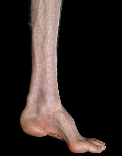
Charcot–Marie–Tooth disease (HSMN 1 and 2) (Figure 2)

  • Autosomal dominant.
  • Motor and sensory demyelinating neuropathy (motor more than sensory defects). 
  • Charcot–Marie–Tooth (CMT) hypertrophic form (HMSN 1) – onset second decade of life. 
  • CMT axonal form (HSMN 2) – onset third or fourth decade, more extensive foot involvement “stork-leg”appearance.
  • Orthopaedic manifestations:
  • Pes cavus
  • Hammer toes
  • Peroneal weakness
  • Stork legs/upside down champagne bottle legs
  • Intrinsic muscle wasting in the hands
  • Investigations –Low nerve conduction velocities in peroneal, ulnar and median nerves.
  • Treatment:
  • Surgery for pes cavus
  • Intrinsic minus procedure for hands

BS3NMD 2.jpg

Fig 2. Charcot Marie Tooth Disease.Signs and symptoms include: weakness in the legs, ankles and feet, frequent falling, numbness and pain in legs, loss of muscle bulk in the legs and feet, hammertoes, high foot arches, decreased ability to walk and run, and footdrop.

Dejerine Sottas disease (HSMN 3)

  • Autosomal recessive.
  • Hypertrophic neuropathy of infancy CMT3.

Clinically:

  • Delayed ambulation
  • Pes cavus
  • Stocking glove dysaesthesia
  • Foot drop
  • Spinal deformities
  • Confined to wheelchair by third/fourth decade.

Riley Day syndrome

  • Autosomal recessive.
  • Sensory and autonomic neuropathies. 
  • Only in Ashkenazi Jews.

Clinically:

  • Dysphagia
  • Alacrima
  • Pneumonia
  • Excess sweating
  • Postural hypotension
  • Sensory loss

Hereditary sensory neuropathy

  • Congenital insensitivity to pain and temperature. 
  • Autosomal dominant or recessive. 
  • Clinically Charcot joints and foot ulceration.

Friedrich’s ataxia

  • Spinocerebellar disorder but may have degeneration of posterior root ganglia and peripheral nerves. 
  • Autosomal recessive.
  • Clinical onset before the age of 10 years.
  • Staggering broad-based gait, nystagmus, ataxia, cardiomyopathy, cavus foot, scoliosis, motor and sensory deficits.
  • Death by 40–50 years.

Poliomyelitis

  • Pathology – viral destruction of anterior horn cell of spinal cord and brainstem. 
  • The virus gains entry via the gastrointestinal tract.
  • A mild ’flu like illness followed by residual paralysis 6 months later is typical. 
  • Causes lower motor neuron weakness plus or minus difficulty in breathing or swallowing.
  • Contagious for 4 weeks.
  • Rare in developed countries following widespread vaccination.
  • Post-polio syndrome can occur many years later due to progressive muscle weakness in both old and new muscle groups due to “neural fatigue.”
  • Principles of treatment are to get the patient walking, prevent deformity, decrease the need for bracing, correct upper limb problems and prevent scoliosis.

Treatment:

  • Rest and isolation acutely
  • Physiotherapy
  • Isolated muscle weakness with no deformity can be treated with a splint or tendon transfer.
  • If there is a deformity surgery can directed to the soft tissue if it is passively correctible, or bones if it is not correctible.

Spinal muscular atrophy

  • Autosomal recessive.
  • Due to the loss of anterior horn cells from the spinal cord. 
  • Most common diagnosis in girlswith progressive weakness. 
  • Normal intelligence. 

Classification: 

  • Type 1 or acute Werdnig–Hoffman form. Presents at birth, short lifespan 
  • Type 2 or chronic Werdnig–Hoffman form. Presents 6 months to 2 years of age 
  • Type 3 or Kugelberg–Wellander form. Presents after 2 years ofage, long lifespan 

Clinically:

  • Progressive scoliosis
  • Symmetric paresis with more involvement of lower limbs and proximal muscles
  • Management:
  • Bracing and wheelchair modifications are useful initially. 
  • Spinal stabilisation is indicated for children over10 years of age with a flexible curve of more than 40 degrees and forced expiratory volume (FEV) more than 40%. 
  • Spinal fusion can result in loss of lordosis which is necessary for walking, thus should be considered when child stops walking to aid sitting and breathing.
  • Hip dysplasia– coxa valgatreated by varus osteotomy with or without acetabular procedures.

Neuromuscular junction – myasthenia gravis

  • Competitive inhibition of acetylcholine receptors at motor end plate by autoantibodies produced by the thymus gland.
  • Insidious onset of muscle fatigability after exercise. 
  • Treatment – cyclosporin, anti-acetylcholinesterase agents, thymectomy.
  • Non-inflammatory inherited disorders with progressive muscle weakness.
  • Treatment is largely physiotherapy, genetic counselling and orthotics.
  • Surgical intervention may take the form of tibialis posterior tendon transfers, release of contractures and early fusion for neuromuscular scoliosis.

Duchenne’s muscular dystrophy

  • Sex linked recessive (boys).

Clinically: 

  • Clumsy walking 
  • Decreased motor skills 
  • 95% get scoliosis 
  • Lumbar lordosis 
  • Calf pseudohypertrophy 
  • Positive Gower’s sign (raises by walking hands up the legs to compensate for gluteus maximus and quads weakness – Figure 3)
  • Hip extensors typically affected first
 
Figure 3.Clinical diagnosis of DMD

Tests:

  • Very high creatine phosphokinase (CPK) 
  • Absent dystrophin on muscle biopsy and DNA testing 
  • Foci of necrosis and connective tissue infiltration on muscle biopsy

Natural history: 

  • Loss of independent ambulation by 10 years, with KAFOS can walk to 12–14 years 
  • Wheelchair bound by 15 years 
  • Scoliosis 13–15 years. Treat with posterior fusion and instrumentation
  • Bedridden aged 16 years 
  • Death by age 20 years from cardiorespiratory complications

BS3NMD 3.png

Figure 4. Gowers sign 

Becker’s dystrophy

  • Sex linked recessive (boys).
  • Reduction in dystrophin.
  • Clinically manifests around the age of 7 years. Less severe than Duchenne’s.
  • Associated with colour blindness.
  • Most live past 22 years without respiratory support.
  • Abnormal dystrophin on muscle biopsy.

Fascioscapulohumeral muscular dystrophy

  • Autosomal dominant.
  • Weakness of face, shoulder girdle and arm muscles.
  • Patients are typically between 6 and 20 years old.

Associated with:

  1. Facial muscle abnormalities
  2. Winging of scapular
  3. Foot drop
  4. Normal life expectancy
  • Normal CPK.
  • Surgery if needed is scapulothoracic fusion.
  • Autosomal dominant disorders.
  • Inability to relax after muscle contraction.
  • Normal enzyme assays, EMG shows characteristic “dive bomber” pattern. 
  • Myotonia congenita chromosome 7, muscle chloride channel problem. 
  • Widespread involvement, no weakness.
  • Dytrophic myotonicachromosome 19, small gonads, heart disease, low IQ, distal, lower limb involvement.
  • Paramyotonia.
  • Chromosome 17, sodium channel problem. Myotonia in cold weather.
  • Definition = congenital non-progressive limitation of joint movement due to soft tissue contractures affecting two or more joints.

Arthrogrypotic multiplex congenita (Figure 5)

  • Non-progressive disease.
  • Multiply congenitally rigid joints.
  • Can be myopathic, neuropathic or mixed. 
  • Associated with a loss of anterior horn cells and other neural elements of the spinal cord.
  • Normal intelligence, no visceral abnormalities and normal facies.
  • Upper limb:
    • Adduction and internal rotation of shoulder
    • Extension of elbow; elbow has no creases
    • Flexion and ulnar deviation of wrist
  • Lower limb:
    • Teratological hip dislocations
    • Knee contractures
    • Resistant clubfeet
    • Vertical talus
  • Spine:
    • Scoliosis in 35%

Multiple aetiologies: 

  • ? Fetal packing
  • ? Intra-uterine event at weeks 8–12
  • ? Intra utero virus 

Treatment:

  • Passive stretching
  • Serial casts
  • Osteotomies
  • One upper limb left in extension, one in flexion
  • In lower limb, soft tissue releases (knee and ankles), open reduction hip dislocation (bilateral dislocations usually left alone)
  • Knee contracture = posterior capsulotomy and hamstring division after feet treated (up to 6 months of age)
  • Latersupracondylar osteotomy and shortening(avoids stretching neurovascular bundle (NVB)) better
  • Scoliosismanaged like idiopathic curves
  • Surgical principle in arthrogryposis = “do no harm”

BSNMD 4.png

Figure 5. Arthrogrypotic Multiplex Congenita

Larsen’s syndrome

  • Similar but fewer joints are less rigid.
  • Associated with multiple joint dislocations, including bilateral knee dislocations.
  • Also flat face, cervical kyphosis, clubfeet, scoliosis.
  • Normal intelligence.

Distal arthrogryposis syndrome

  • Autosomal dominant.
  • Mainly affects the hands and feet.
  • Ulnarly deviated fingers at metacarpophalangeal (MCP) joints.
  • MCP and proximal interphalangeal joint flexion contractures.
  • Adducted thumbs with web space thickening.
  • Clubfoot and vertical talus often.

Multiple pterygium syndrome 

  • Autosomal recessive.
  • Cutaneous flexor surface webs at knee and elbow.
  • Congenital vertical talus, and scoliosis.
Previous
Next

QUESTION 1 OF 2

38.You review a 19-year-old male patient in outpatients who has suffered a progressive muscle weakness and has been declining recently. He has a waddling gait and positive Gower’s sign. He walks on his tiptoes and you not calf enlargement.
Which investigation is the best to confirm your diagnosis?

QUESTION ID: 2226

1. Creatine Kinase
2. EMG
3. Genetic testing
4. MRI Calf
5. Muscle biopsy and immunohistochemistry

QUESTION 2 OF 2

110.Concerning the neuromuscular junction(motor end plate)

QUESTION ID: 2245

1. Botox blocks AC receptors at the motor end plates
2. Malignant hyperthermia is a life-threatening clinical syndrome of hypermetabolism involving skeletal muscle due to an allergy
3. Neostigmine is an antidote for non-depolarizing blockers
4. Succinylcholine blocks the effect of AC at the neuromuscular junction
5. With Myasthenia gravis the release of AC from the end plate is blocked