TOPIC DETAILS

  Team Member Role(s) Profile
Paul Banaszkiewicz Paul Banaszkiewicz Section Editor
James Donaldson James Donaldson Segment Author

The two terms, “osteochondritis” and “osteochondrosis,” are sometimes confused.

  • The osteochondroses are a heterogeneous group of disorders affecting epiphyses, physes, and apophyses of children.
  • They are generally self-limiting diseases characterised initially by avascular necrosis (AVN) of primary or secondary centres of ossi?cation, followed by spontaneous healing.
  • The aetiology is not fully understood, but vascular compromise, disorders of coagulation, and genetic/hereditary causes have been implicated.

The osteochondroses belong to three groups:

Those involving articulations:

  • Perthes disease affecting the femoral head.
  • Kohler’s disease affecting the navicular.
  • Freiberg’s disease affecting metacarpal heads (usually the second).
  • Kienbock’s disease affecting the lunate.

Those involving tendon entheses (bone attachments):

  • Osgood Slater’s disease.

Those affecting the spine:

  • Scheuermann’s disease
  • Osteochondritis dissecans (OCD) – this is a form of osteochondral fracture or an epiphyseal osteochondrosis. It is sometimes included among the osteochondromatoses as the disorder is characterised by avascular necrosis. However, it differs from the osteochondromatoses in that adults may also develop the disorder and loose bodies commonly develop.
  • Occurs at an epiphysis at the musculotendinous junction.
  • Acute or chronic trauma and genetics play a role in aetiology.

Osgood Schlatter’s disease (OSD) (Figure 1)

  • Common cause of knee pain in adolescent boys aged 9–13 years.
  • Presents with pain and swelling over tibial tubercle.
  • Gradual onset. Worse with activity and eased with rest.
  • Pain with resisted extension.
  • Tender over tibial tubercle.
  • Normal knee exam.
  • May be associated with hamstring tightness.
  • Bilateral in 20–50%.
  • 50% may have a history of trauma.
  • The most commonly accepted theory is that repeated traction through the patella tendon on the anterior portion of the developing ossification centre leads to multiple subacute fractures or tendinous inflammation.
  • Self-limiting. 90% resolve within 1 year.
  • After maturity an unfused ossicle or overlying bursa may continue to cause pain.

BS3OSTEOCHONDRITIDES 1.jpg

Figure  1. Osgood Schlatter’s disease

Sinding–Larsen–Johansen syndrome (Figures 2 and 3)

  • Similar to OSD but the pain and radiographic changes are localised to the lower pole of the patella.
  • Caused by increased tension and pressure from repetitive traction by the patellar tendon on the lower pole of the immature patella.
  • Treatment is rest.

BS3OSTEOCHONDRITIDES 2.jpg

Figure 2.  Lateral knee radiograph Sinding-Larsen-Johansen syndrome 

BS3OSTEOCHONDRITIDES 3.jpg

Figure 3. MRI Sinding-Larsen-Johansen syndrome

  • Is a painful inflammation of the calcaneal apophysis.
  • Presents with heel pain in a growing, active child
  • The aetiology is believed to be due to repetitive trauma to the weaker structure of the apophysis, induced by the pull of the tendo Achilles on its insertion. During the rapid growth surrounding puberty, the apophyseal line appears to be weakened further because of increased fragile calcified cartilage.
  • A lateral radiograph excludes other causes of heel pain.
  • The presence of fragmentation and sclerosis radiographically is of questionable significance.
  • Treatment involves reduction in activity and occasionally a plaster.

Epiphysitis of fifth metatarsal base (Figures 4 and 5) AKA Iselin’s disease

  • Presents with pain and prominence at the fifth metatarsal (MT) base in a child.
  • More common in girls.
  • Due to repetitive traction of the peroneus brevis tendon attachment.
  • Treated with restricted activity and occasionally a plaster.

BS3OSTEOCHONDRITIDES 4.jpg

Figure 4. Radiograph epiphysitis of 5th metatarsal base

BS3OSTEOCHONDRITIDES 5.jpg

 Figure 5. MRI Epiphysitis of 5th metatarsal base

  • May present with groin pain and restricted function.
  • Treated with rest and anti-inflammatories.
  • van Neck Odelberg disease.
  • Presents with groin pain in a child 9–12 years old.
  • Radiographs show irregularity and cystic changes over ischiopubic synchondrosis.
  • Likely to be due to traction from the insertion of the hip adductors or extensors.

Perthes disease

  • Idiopathic AVN of proximal femoral epiphysis.
  • See “paediatrics.”

Kohler’s disease (Figures 6 and 7)

  • Avascular necrosis of navicular bone.
  • Unclear aetiology.
  • Usually children 4–7 years old.
  • Boys > girls.
  • Can be bilateral in 25%.
  • The blood supply of the central one third of the navicular is a watershed zone which accounts for the susceptibility to AVN (and stress fractures).
  • The navicular is the last bone to ossify, which also increases its vulnerability to mechanical compression and injury.
  • Typically a self-limiting condition for 1–3 years after diagnosis.
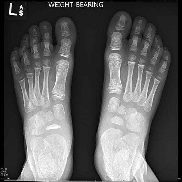
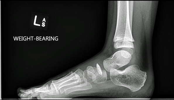
  • Radiographs show characteristic clerosis, fragmentation, and flattening of the navicular.
  • Most tarsal navicular bones reorganise after the disease has run its course; some continue to be deformed but remain largely asymptomatic.
  • Treated with rest, anti-inflammatories and occasionally a walking cast.
BS3OSTEOCHONDRITIDES 6.png

 Figure 6. Weight bearing radiographs Kochler's disease
BS3OSTEOCHONDRITIDES 7.png 

Figure 7. Lateral radiograph Kochler's disease

  • Freiberg’s disease (Figure 8)
  • AVN of second metatarsal head.
  • Commonly in adolescents.
  • Girls > boys.
  • More common with a long second MT.
  • Presents with localised pain, swelling and restricted movement.
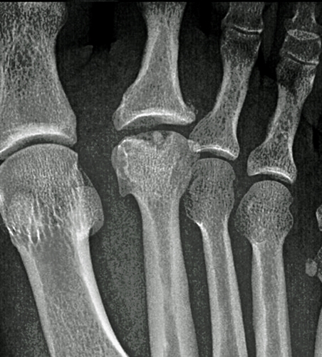
  • Radiogrpahs show a progression of sclerosis to flattening of the MT head to collapse and joint destruction in later disease.

Smillie classification – grades 1–5:

1.     Magnetic resonance imaging diagnosed subchondral fracture

2.     Dorsal collapse on X-ray

3.     Collapse of dorsal MT head

4.     Collapse of entire head with joint space narrowing

5.     Secondary osteoarthritis

·       Initial conservative Mx as above.

·       Surgery may involve a dorsiflexion closing and shortening osteotomy or partial MT head resection.
BS3OSTEOCHONDRITIDES 8.jpg

 Figure 8. Radiograph Frelberg's disease

Osteochondritis dissecans (Figures 9–11)

  • Characterised by a separation of an osteochondral fragment from the articular
  • The underlying bone from which the fragment separates has normal vascularity and this separates OCD from osteonecrosis.

Approximately 75% cases affect 10–20 year olds:

  • Juvenile form that affects the skeletally immature with open growth plates.
  • Adult form that affects age range 15–50 years in which growth plates are fused.
  • Occurs in the knee 70% of the time; in 75% of knee cases it involves the lateral aspect medial femoral condyle
  • 6% of cases in capitellum elbow, 4% in talus.
  • Males > Females 2:1.
  • Bimodal distribution involving adolescents and young adults.

Aetiology unclear, likely multifactorial:

Trauma:

  • Indirect or repetitive microtrauma most likely.
  • Persistent microstresses when the bone is growing and the blood supply somewhat tenuous interrupts the cascular supply leading to subchondral bone infarction.
  • Direct trauma may account for medial talar lesions.

Ischaemia:

  • Unlikely/controversial.
  • Little objective evidence for a genetic or endocrine cause although the condition may run in families.

BS3OSTEOCHONDRITIDES 9.jpg

BS3OSTEOCHONDRITIDES 10.jpg

BS3OSTEOCHONDRITIDES 11.jpg

Figures 9- 11. Radiographs Osteochondritis Dissecans

Pathophysiology

  • Wedge-shaped area of dead tissue typical of an infarct due to poor blood flow through a single end artery.
  • The arteries supplying blood to subchondral bone are end arteries and therefore the infarction occurs in the distribution of one or a small number of end arteries.
  • The infracted bone is surrounded by viable bone. The presence of dead bone initiates a reaction that includes osteoclastic resorption of the necrotic tissue and the formation of a fibrous tissue shell around the piece of necrotic bone.
  • The overlying cartilage receives nutrition from the synovial fluid and is therefore viable in the early stages of the disease.
  • Alterations in the balance between cartilage matrix synthesis and breakdown secondary to altered nutrition renders the cartilage soft and spongy.
  • In the juvenile form the process may reverse with healing of the bone infarction; however, in the adult form the process of walling off usually continues.
  • Bone resorption leads to a flattening of the contour of the articular surface and eventually the dead bone and its attached viable cartilage may break away from the underlying bone under load, forming an osteochondral loose body and a defect in the articular cartilage. This is usually 1–2cm in diameter and around 1 cm deep.
  • There is potential for the defect to heal with fibrocartilage if the floor is damaged and stem cells from the bone marrow proliferate, but in general the defect remains unhealed leading to altered biomechanics and secondary osteoarthritis.

·       Typically four stages:

  • Stage I consists of a small area of compression of subchondral bone.
  • Stage II consists of a partially detached osteochondral fragment. A radiograph of the bone may reveal a well circumscribed area of sclerotic subchondral bone separated from the remainder of the epiphysis by a radiolucent line.
  • Stage III lesions are the most common and consist of a completely detached fragment that remains within the underlying crater bed.
  • Stage IV lesions consist of a completely detached fragment that is completely displaced from the crater bed. This is also termed a loose body.

Symptoms

  • Vary with stage.
  • Possible history of trauma in approximately50% cases of the juvenile form.
  • Pain, swelling, locking, giving way, catching and grinding.
  • Formation of a loose body may give rise to dramatic secondary symptoms.
  • Clinical findings may include an effusion, crepitus and localised tenderness.
  • For MFC lesions lateral aspect patients may walk with the leg externally rotated to prevent spinous process impinging on the lateral aspect medial femoral condyle (MFC). Wilson’s test positive.

·       Surgery if:

  • symptoms in a child for more than 6–12 months
  • inadequate healing radiographically
  • loose body present
  • skeletal maturity expected within 6 months.
  • Usually earlier surgery in an adult as healing is unlikely.

Investigations

Plain radiographs

  • May be normal in very early stages. Luciencies in the epiphysis and loss of definition of the usually sharp cortical line of the subchondral bone of the joint surface. If lesions are visible:
  • Stage I: normal radiograph.
  • Stage II: partially detached osteochondral fragment.
  • Stage III: complete, non-displaced fracture remaining within the bony crater.
  • Stage IV: detached and loose osteochondral fragment.

Computed tomography

  • Better able to demonstrate the lesion compared to radiographs.

Magnetic resonance imaging

  • Useful and allows preoperative planning:
  • Stage I: bone marrow oedema.
  • Stage IIa: subchondral cyst.
  • Stage IIb: incomplete separation of the osteochondral fragment.
  • Stage III: fluid around an undetached, undisplaced osteochondral fragment.
  • Stage IV: dispalced osteochondral fragments.

Bone scan

  • The higher the osseous uptake the more potential for healing.

Arthroscopic examination of the joint

  • This can reveal information about the status of the cartilage, alterations in the contour of the articular surfaces and the presence of loose bodies. Arthroscopic changes are progressive.
  • Articular cartilage is smooth and intact but may be soft or ballottable.
  • Articular cartilage has a rough surface.
  • Articular cartilage has ?brillations or ?ssures.
  • Articular cartilage with a ?ap or exposed bone.
  • Loose, non-displaced osteochondral fragment.
  • Displaced osteochondral fragment.

Treatment

  • Conservative for children with no loose bodies:
  • Protected weight-bearing.
  • Limited activities.
  • Surgical options include:
  • Arthroscopic subchondral drilling.
  • Arthroscopic debridement and fragment stabilisation (Figures 12 and 13).
  • Arthroscopic excision, curettage, and drilling.
  • Open removal of loose bodies, reconstruction of the crater base, mosaicplasty for fragment healing, and potential replacement with fixation.
  • Autologous chondrocyte transplantation.
  • Radical removal of sclerotic bone with bone grafting of the defect and autologous chondrocyte transplantation (i.e. sandwich technique).

BS3OSTEOCHONDRITIDES 12.gif

BS3OSTEOCHONDRITIDES 13.gif

Figures 12- 13. MRI demonstrating internal fixation of undisplaced lesions of osteochondritis dissecans in the knee

Prognosis

  • Juvenile OCD without a loose body usually heals (94%)
  • Adult OCD is unlikely to heal and secondary osteoarthritis in the medium to long term is more likely.
Previous
Next

References

  • 1. Freemont T. Osteochondritis. Orthopaedics and Trauma 2010; 24(6):410-441