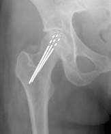
TOPIC DETAILS

  Team Member Role(s) Profile
Paul Banaszkiewicz Paul Banaszkiewicz Section Editor
James Donaldson James Donaldson Segment Author
  • Death of bone from ischaemia (not disease).
  • Also known as avascular necrosis (AVN) but osteonecrosis (ON) is preferred as it is a correct description of the histopathological process and does not suggest a specific aetiology. Osteonecrotic bone is NOT avascular, vessels are present but compromised.
  • Osteocytes will survive 12–48 hours of ischaemia and marrow cells 6 hours.
  • Average age is 35–50 years.
  • Slight male preponderance.
  • Most commonly involves the femoral head, then in descending order: medial femoral condyle of the knee, humeral head, talus, lunate, capitellum and metatarsal heads.
  • In the hips it is bilateral 80% of the time.
  • Three per cent may have multifocal involvement (more than three sites involved).
  • Five per cent of total hip arthroplasty (THA) performed as a result of osteoarthritis (OA) secondary to AVN.
  • Insidious pain which starts initially with activity, then at rest and eventually at night.
  • Pain may be severe for 6–8 weeks and then subside.
  • Mild symptoms and effusion usually persists.
  • Night pain may be an early feature.
  • Pain may be present for many months before X-ray changes are manifest.
  • Earliest examination finding is decreased internal rotation.

Idiopathic (40%):

  • May be an unidentified coagulation abnormality 

Arterial insufficiency:

  • Fracture 
  • Dislocation 
  • Infection 

Arteriolar occlusion:

  • Sickle cell disease and haemoglobinopathies 
  • Caisson disease in divers and compressed air workers (e.g. miners)
  • Vasculitis, e.g. systemic lupus erythematosus, irradiation 

Capillary occlusion:

  • Fatty infiltration due to steroids, alcohol and other drugs, e.g. chemotherapy 
  • Systemic steroid treatment (37%)
  • High alcohol intake (20%) 
  • Gaucher’s disease
  • Hyperlipidaemia
  • Venous occlusion 

Other:

  • Renal transplant patients (16% will develop ON) 
  • Pancreatitis 
  • Haematological malignancies, e.g. lymphomas, leukaemias 
  • Diabetes mellitus 
  • Endotoxin reactions 
  • Toxic shock 
  • Inflammatory bowel disease 
  • Brain/spinal surgery 
  • Anticoagulant deficiencies 
  • Nephrotic syndrome

Intraosseous hypertension: 

  • Raised intraosseous pressures have been consistently found in cases of AVN.
  • Blood flow through the intraosseous compartment is inversely proportional to the bone marrow pressure.

Abnormal extraosseous blood flow:

  • Superselective angiography of the medial femoral circumflex artery has been used for study.
  • There is a consistent loss of transcortical blood flow from the superior retinacular arteries and alterations in the revascularisation process in hips with radiographically and preradiographically defined osteonecrosis.
  • A mechanism of alteration of the process of revascularisation has been considered to be a contributory feature of non-reversible osteonecrosis.

Fat embolism:

  • Fatty osteocytic necrosis is felt to progress to ischaemic degeneration of necrotic osteocytes and adipocytes.
  • When the volume of subchondral fat overload results in vascular stasis, local hypercoagulability, endothelial damage, and subsequent intravascular coagulation.
  • In this situation, local mechanisms of repair are prevented.
  • Histologically observed phenomena include intraosseous thromboses and peripheral haemorrhages. These phenomena were observed in animals with induced intravascular coagulopathy and in children with disseminated intravascular coagulopathy and histological evidence of interosseous thrombosis and osteonecrosis. 

Fat cell abnormalities: 

  • Fat cell hypertrophy and fatty marrow overload have been demonstrated in animals as a consistent consequence of high-dose corticosteroid exposure.
  • Histological changes consistent with necrosis were frequently identified and femoral head blood flow was consistently diminished,
  • The use of lipid clearing agents was shown consistently to improve this alteration in blood flow. 
  • The earliest microscopic signs indicative of bone ischaemia are seen in the marrow spaces, where starting from the second day there is loss of nuclear staining of marrow cells and large round and ovoid spaces filled with fat appear.
  • The fatty and haemopoietic marrow then becomes ghosted and the small vessels show evidence of necrosis.
  • Necrotic osteocytes may appear normal for weeks by light microscopy.
  • After 15 days the osteocytic lacunae are empty and the trabecular surface is devoid of cells. 
  • At the border of the necrotic zone there is proliferation of capillaries accompanied by fibroblasts and foamy histiocytes, which are responsible for the breakdown of necrotic fatty marrow, while dead bone is partly removed by osteoclasts and substituted by newly formed trabeculae; alternatively woven bone is laid down on the surface of dead trabeculae.
  • Absence of remodeling may cause the development of stress fractures.
  • Repair and revascularisation advances from the vascular margin causing appositional new bone and bone resorption.
  • Dead bone may fracture and collapse or repair maintaining joint integrity and congruency.

BS3ON 1 .jpg

Figure 1. AP radiograph hip osteonecrosis 

BS3ON 2 .jpg

Figure 2. MRI talar neck ON

  • Bone death.
  • Inflammation/revascularisation:
  • Early in the course of healing of ON, there is a reactive hyperemia. Vascular fibrous repair tissue proliferates adjacent to bone. The dead bone becomes walled in by this tissue in a fashion similar to sequestrum formation in osteomyelitis.
  • Repair:
  • Revascularisation of the dead bone takes several months to years. Cutting cones carrying blood vessels enter the dead bone. Dead bone is removed by osteoclastic resorption. New bone is laid down on top of the dead trabeculae by osteoblasts– Phemister coined the term “creeping substitution” to indicate this slow replacement of aseptic dead bone in contrast with the massive resorption or sequestration of dead bone associated with osteomyelitis. 
  • Remodeling:
  • At the margin of the creeping zone of substitution with viable vascularised bone, osseous reinforcement occurs in a compensatory response to bone weakening caused by the reactive interface. This progresses to a rim of sclerosis that is frequently undulating or serpentine in morphology. The reactive interface undergoes progressive remodeling and repair at the junction with the area of ON. Unfortunately, this creeping zone of substitution neither creeps nor substitutes, nor repairs the areas of ON extensively in the vast majority of cases.
  • Intact articular cartilage except at the edge of necrotic area, which exhibits cracking and folding.
  • Necrotic area in cross-section is yellow, opaque, chalky with hyperaemic fibrous tissue at the margin(Figure 3).
  • Adjacent bone may be thickened.
  • Late changes are breaks in smooth contour of the bone, destruction of articular cartilage, loose bodies and marginal osteophytes (secondary osteoarthritis).

BS3ON 3 .jpg

Figure 3. Histological specimen of AVN femoral head

BS3ON 4 .jpg

Figure 4. Photomicrograph of a histologic preparation of a femoral head with ON.

  • Photomicrograph of a histological preparation of a femoral head with ON(Figure 4). The thickened trabeculae of the viable bone at the base of the infarct can be clearly appreciated (H&E staining).
  • Dead trabeculae (empty lacunae) stain deeper blue than non-necrotic bone and have ragged margins with osteoclasts on one side and osteoblasts on the other.
  • Lacunae may be enlarged and cystic or normal size with pyknotic nuclei.
  • Marrow has fat necrosis and calcium deposits (marrow is a more sensitive indicator of necrosis than bone).

BS3ON 5 .jpg

Figure 5. Photomicrograph of stage III ON.

  • Photomicrograph through an area of stage III osteonecrosis(Figure 5). 
  • Demonstrates viable articular cartilage as well as the necrotic bone and bone marrow of the infarcted area; between them is the subchondral crescent.
  • The lower portion of the frame shows the junction between the necrotic and viable bone. 
  • At the bottom of the picture the thickened trabeculae of viable bone are evident. This thickening is the result of new bone deposition on the trabecular surfaces, which occurs as part of the healing of the infarct.

BS3ON 6 .jpg

Figure 6. Fat cell necrosis and fibroblastic proliferation at margin infracted area

  • Photomicrograph to demonstrate focal fat necrosis as well as fibroblastic and vascular proliferationat the margin of the infarcted area(Figure 6).

X-ray

  • Normal in stages 0 and I.
  • Osteopenia and sclerosis in stage II.
  • Subchondral collapse "crescent sign" in stage III.
  • Secondary OA in stage IV.

Magnetic resonance imaging (MRI)

  • Ischaemic marrow changes are evident before bone changes are apparent.
  • Earliest finding:
  • T1 images – decreased signal from ischaemic marrow/single band-like area of low signal intensity.
  • T2 images a second, high signal intensity line can be found within the line seen on T1 images, believed to represent hypervascular granulation tissue ("double line sign").
  • Rarely AVN may be found on histology with a normal MRI.
  • 100% sensitivity, 98% specificity.

Shimizu, et al. Prediction of collapse with MRI of AVN of the femoral head. JBJS 1994; 76A.

  • Where at least three-quarters of the diameter of the femoral head and at least two-thirds of the major weight-bearing area is involved – 74% had collapsed within 32 months.
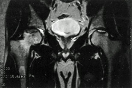
BS3ON 7.png

Figure 7. Bilateral hip AVN on coronal MRI.

Bone scan

  • Cold area may only be evident in early stages of disease (14–21 days) prior to revascularisation.
  • Usually increased uptake at time of study.
  • Increased uptake on both sides of joint suggests OA rather than AVN.
  • 75–80% sensitivity in precollapse stage.

Single photon emission computed tomography (SPECT) scan

  • SPECT imaging is a three-dimensional isotope scanning technique.
  • It has been shown to be useful in the analysis of bone graft healing and in the identification of early osteonecrosis.
  • Because of its ability to visualise bone reactivity in three dimensions, it is helpful in identifying an area of decreased activity (cold spot) within an area of increased activity.
  • This clinical picture can be seen in a very early stage of osteonecrosis.

Classic reference1

Ficat RP. Idiopathic bone necrosis of the femoral head. Early diagnosis and treatment. J Bone Joint Surg Br 1985; 67(1): 3–9.

  • Ficat and Arlet proposed the original classification of osteonecrosis in 1964 before the advent of MRI. It consisted of stage I through to stage IV and did not include stage 0.
  • This article in 1985 modified the original classification (1964) by including functional exploration of bone and adding a stage 0 previously identified by Hungerford in 1979.2
  • Later on at least three further changes to this modified Ficat system have occurred to take account of MRI findings, patient symptoms and modifications of original radiographic findings described.3From having three stages, the system progressed to four stages and then up to six stages, sometimes including symptoms and MRI findings.3Most surgeons still use the simple four-tiered method.
  • Despite Mont et al.3identifying at least 16 classification systems in use to grade and describe avascular necrosis, the Ficat system continues to be the most widely used system. One significant change is that histological examination of the hip (biopsy) is no longer needed for a diagnosis of AVN. MRI is the most sensitive and specific diagnostic method.
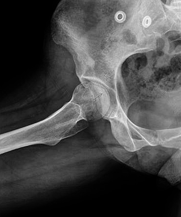
BS3ON 8.jpg

Figure 8. Crescent sign on a lateral hip X-ray
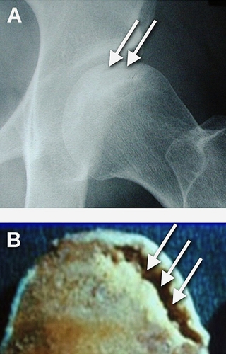
BS3ON 9.jpg

Figure 9. Nontraumatic Osteonecrosis of the Femoral Head

Ficat classification

The Ficat classification is a five-stage system:

  1. Preclinical: known as a silent hip. No clinical symptoms. Normal radiographs. MRI non diagnostic.
  2. Pre-radiographic: radiographs usually normal or at most show only minor changes such as subtle loss of clarity with poor definition or blurring of trabeculae pattern. Diagnosed with a positive MRI or bone scan. Earliest clinical manifestation syndrome. Usually presents with sudden onset of hip pain.
  3. Pre-collapse (before flattening of the head or sequestrum formation): this extends over several months with clinical symptoms and signs persisting or worsening. Radiographs demonstrate osteopenia/sclerosis of the femoral head, spherical head, crescent line due to subchondral fracture.
  4. Collapse: segmental flattening and collapse of the femoral head. Worsening pain, limp, limited range of motion in all planes.
  5. Osteoarthritis: terminal phase, secondary degenerative change superimposed on a deformed femoral head.
  • Stages 0 to II were described as early stages and stages III and IV were classified as late stages. 

Steinberg modification of Ficat classification:4

  • Grade 0
  • Normal imaging
  • Preclinical
  • Grade 1
  • Normal X-ray
  • Early abnormal MRI changes
  • Grade 2
  • Osteopenia and sclerosis on X-ray
  • Grade 3
  • Crescent sign
  • Grade 4
  • Flattening of the femoral head
  • Grade 5
  • Joint space narrowing
  • Grade 6
  • Secondary OA
  • Stages I–V can be further subdivided into mild (A), moderate (B) or severe (C) to include the extent of femoral head involvement.

Natural history5

  • Meta analysis of the literature: 21 studies involving 819 hips, average follow-up 34 months, all treated non-operatively (various protocols of weight-bearing status)
  • Rates of preservation of the femoral head: 
 

Stage 1

35% 

Stage 2

31% 

Stage 3

13% 

Treatment6

  • Potentially reversible early if corticosteroids or alcohol stopped.
  • Symptomatic treatment, weight loss and physiotherapy.
  • Bisphosphonates may be of benefit in the pre-collapse stages (controversial).
  • Protective weightbearing: 
  • Start with non-weightbearing with progression to weightbearing allowed as clinical symptoms and signs demonstrate that the hip is less irritable. 
  • Radiographic and clinical follow-up at 6 week intervals until pain has subsided.
  • Success 5–20% at 3–5 years follow-up.
  • Core decompression: 
  • The goal of this procedure is to demonstrate intraosseous venous hypertension and remove a central core of bone from the lesional area effectively to lower intraosseous pressure.
  • The biopsy obtained can confirm the disease histologically.
  • Patients remain non-weightbearing for 6 weeks post-op.
  • There has been substantial controversy as to the effectiveness of this procedure.
  • Meta-analysis of 24 studies involving 1206 hips treated with core decompression.
  • Rates of preservation of the femoral head:


Core decomp.

No Rx

Stage 1

84%

35% 

Stage 2

65%

31% 

Stage 3

47%

13% 


BS3ON 10.png

Figure 10. Core decompression

BS3ON 11.png

Figure 11. Drill lines demonstrate the pathway of small drill holes used in core decompression

  • Electrical stimulation: 
  • The use of pulsed electromagnetic field therapy with external coils.
  • Not commonly used.
  • Proximal femoral osteotomy:
  • This may compromise later total hip replacement (THR). 
  • The procedure directly addresses the mechanical aspects of osteonecrosis on the femoral head.
  • The goal is preservation of the femoral head by altering the pattern of stress transfer in the diseased head.
  • Varus or rotational osteotomies may be used.
BS3ON 12.gif

Figure 12. Radiograph of proximal femoral osteotomy
  • Strut grafting (fibula/tibia/iliac crest)\;
  • This procedure, either of the Bonfiglio non-vascularised type or using a vascularised grafting technique, may be used in the pre-collapse stages.
  • Cortical strut grafts, e.g. ilium, tibia, fibula, are placed into a core track in the femoral neck under the subchondral bone to help prevent collapse.
  • It does not alter the stress-transfer patterns in the upper femoral regions.
  • Although early reports of vascularised grafting techniques were encouraging, studies to date are not sufficient to support widespread application(Figure 13).
BS3ON 13.jpg

Figure 13. Vascularised strut grafting

Vascularised pedicle flaps: 

  • Quadratus femoris graft (metres) – posterior.
  • Tensor fascia lata muscle – anterior.
  • Rarely used.

Trapdoor procedure:

  • Indicated for pre-collapse.
  • The surgical procedure includes anterolateral dislocation of the femoral head to expose the area of the femoral head collapse.
  • The break in the articular cartilage is identified and opened like a trapdoor.
  • Necrotic bone under the flap is excavated and removed. 
  • This defect is filled with cancellous bone graft from the patient’s iliac crest and overpacked into the defect to prevent subsidence. 
  • The flap is then carefully repositioned and the hip reduced and the capsule repaired.
  • The operation is often followed by an acetabuloplasty with or without a varus femoral osteotomy.
  • Good results reported in two studies:

Mont MA, Einhorn TA, Sponseller PD, Hungerford DS. The trapdoor procedure using autogenous cortical and cancellous bone grafts for osteonecrosis of the femoral head. J Bone Joint Surg Br 1998; 80(1): 56–62.

Ko JY, Meyers MH, Wenger DR. “Trapdoor” procedure for osteonecrosis with segmental collapse of the femoral head in teenagers. J Pediatr Orthopaed 1995; 15(1): 7–15.

Advanced stages

Arthrodesis: 

  • More historical, e.g. young patient with unilateral disease.
  • 50–80% of cases are, however, bilateral.

THA: 

  • Both uncemented and cemented total hip arthroplasty have been used in this population.
  • Reported success rates are below what appear to be expected from series in other patient populations.
  • Results are, however, more predictable in terms of relieving pain and function than many of the other options, and many surgeons will replace the hip if symptoms are severe enough with radiographic collapse.
Previous
Next

QUESTION 1 OF 1

A 39-year-old male presents to the orthopaedic clinic with severe acute onset of right hip pain of 8 weeks duration. You suspect osteonecrosis. A medical student in clinic has heard this condition is due to a vascular disturbance in the femoral head blood supply like a compartment syndrome of bone
The most likely vascular disturbance attributed to a compartment syndrome of bone would be 

QUESTION ID: 1243

1. Extraosseous arterial factors
2. Extraosseus extravascular (capsular) factors
3. Intraosseous arterial factors.
4. Intraosseous extravascular factors
5. Intraosseous venous factors

References

  • 1. Banaszkiewicz PA. Idiopathic bone necrosis of the femoral head. Early diagnosis and treatment, in Classic Papers in Orthopaedics. Springer, 2014; 121–123.
  • 2. Hungerford D. Bone-marrow pressure, venography, and core decompression in ischemic necrosis of the femoral-head. Orthopade 1980; 9(4): 245–254.
  • 3. Mont MA, et al., Systematic analysis of classification systems for osteonecrosis of the femoral head. J Bone Joint Surg Am 2006; 88(suppl 3): 16–26.
  • 4. Steinberg ME, Hayken G, Steinberg D. A quantitative system for staging avascular necrosis. J Bone Joint Surg 1995; 77(1): 34–41.
  • 5. Mont MA, Hungerford DS. Non-traumatic avascular necrosis of the femoral head. J Bone Joint Surg Am 1995; 77(3): 459–474.
  • 6. Lieberman JR, et al. Osteonecrosis of the hip: management in the 21st century. Instructional Course Lectures 2002; 52: 337–355.