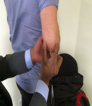
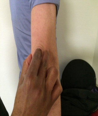
Palpate painful bit last Lateral to medial : examiner should put one finger on the medial epicondyle, one finger on the lateral epicondyle and one onthe tip of the olecranon process.
Lateral
- Palpation should be systematic, involving the lateral supra condylar ridge, lateral epicondyle, common extensor origin, radio-capitellar joint, radial head and lateral collateral Ligament.The extensor carpi radialis brevis and longus muscle can be assessed by resisted wrist extension in neutral and radial deviation, respectively
Differential diagnosis of lateral
elbow pain and swelling
-Tennis elbow
-Radio-capitellar OA
-Osteochondritis dissecans
-Congenital or traumatic radial head
dislocation
-PLRI
Medial
- Tenderness over medial eicondyle and common extensor origin, will indicate medial epicondylitis.The ulnar nerve should be palpating and it can be easily felt behind the medial epicondyle. It is important to identify the position of the ulnar nerve during flexion and extension of the elbow and feel for a subluxing ulnar nerve. If the ulnar nerve is subluxing,it may give rise to medial elbow pain.
Differential diagnosis of Medial elbow pain
Cubital tunnel syndrome
Golfer's elbow
Pronator syndrome
Anterior
Examination of the arm may reveal a retracted biceps muscle bulge appearing more proximally in the arm as opposed to distal muscle bulge with long head of biceps rupture. The structures palpated anteriorly from lateral to medial aspect include brachioradialis, the biceps tendon, brachial artery and the median nerve.The hook test will demonstrate the integrity of the distal biceps tendon at the anterior aspect of the elbow.(Fig 6)

Figure 6. Distal biceps tendon rupture can be demonstrated with 'HOOK TEST'.Ask the patient to actively supinate the forearm against resistance and hook around the biceps insertion from the lateral side.
Posteriorly
- Feel for symmetry of the elbow by feeling these bony prominences. In extension normally these 3 bony prominences form a straight line and in 900degrees of flexion form an isosceles triangle. (Fig 7 and Fig 8) Any abnormality of the above bony landmark arrangement is indicative of previous bony injury.


Figure 7 and Figure 8.The bony relationship of the tip of the olecranon process to the medial and lateral epicondyle with elbow flexed to 900 forms a isosceles triangle and with the elbow extended these bony land marks form a straight line