TOPIC DETAILS

  Team Member Role(s) Profile
Fazal Ali Fazal Ali Section Editor
Caroline Caroline Blakey Segment Author

Document title: Clinical examination of the Child

Section editor: Mr Fazal Ali

Segment author: Miss Caroline Blakey

Acknowledgements: Mr Saif Salih, Miss Hana Salih and Miss Eden Alexander

Document history: Version 1

Introduction

Paediatric orthopaedic examination requires particular skill and takes practice. Where a child is anxious focal examination should be all that is required to confirm a well taken history but remember the adolescent may be modest and reluctant to share symptoms.  Techniques to reassure both the child and the parents require good communication and patience.

To make the younger child feel at ease ensure a friendly environment with toys and space to play.   

A good clinical history is the key to obtaining early diagnosis. It should be sought both from the child and parents in addition to any other adults involved in care. 

The age of the child in relation to the presenting complaint offers common diagnostic differentials.  Specific symptoms such as pain, deformity, gait abnormalities and weakness, should be quantified and ordered chronologically.  With school age children, enquiring whether they can keep up with their peers can give helpful clues.  With younger children knowledge of normal developmental milestones is critical in excluding neuromuscular delay.

Normal developmental milestones indicate the age by which 90% of children would have achieved that particular milestone.

Age       Gross motor skills  Fine motor skills  Social skills 
3 months             Lifts head when prone   Vocalizes without crying, smiles responsively
6 months Sits with support, steady head when sitting Reaches for objects Laughs 
9 months   Sits without support, pulls to stand    ‘dada’, ‘mama’, non-specific
12 months Walks with one hand support    
2 years Runs forwards  Build tower of cubes   Three word sentences
3 years Jumps in place  Dresses self  
4 years Balance on 1 leg    
5 years Hops        Counts 10 objects correctly
6 years Skips Does small buttons, ties laces  


Table 1.Normal developmental milestones indicate the age by which 90% of children would have achieved that particular milestone.

Birth history

Problems during pregnancy or with birth can be associated with both neuromuscular complications and congenital defects.  Intrauterine positioning may be a risk factor for the so-called ‘packaging disorders’.  It is important to elicit concerns during pregnancy regarding fetal movements, history of infection or bleeding.  

Family history

Many paediatric conditions are familial, ranging from generalized  skeletal dysplasias to more focal deformities such as flexible flat feet. Pes cavus in members of the same family suggests a possible hereditary sensorimotor neuropathy.  A positive family history is a strong risk factor for conditions such as developmental hip dysplasia.

Included in the family history is enquiry of siblings, ages and any medical problems.  These questions also give an insight into social circumstances and may highlight areas where families are struggling. 

Systemic Review

The history is completed with review of associated symptoms, along with previous trauma, illness or admissions to hospital, medications and allergies. 

Global examination

The examination itself relies heavily on observation followed by focused examination according to the age of the child and differential diagnosis gained from the history. 

Interacting with the child using toys or props is often more helpful than delivering instructions, particularly with younger children. 

Undress where appropriate but avoid causing embarrassment.  Toddlers and babies can be examined on the parents lap.

General inspection including that of the parent will give clues to syndromic conditions or skeletal dysplasia.  Stature, asymmetry, deformity and facial features should be noted. Look for scars from previous surgery.  Look also for skin and vascular markings as in neurofibromatosis or Klippel-Trenaunay syndrome and note any deficiencies in the limbs including wasting of musculature.  Carefully examine for external stigmata of dysraphis

Clinical Paeds 1.png

Figure 1. Café au lait spots

Standing examination

The height of the child is very informative.  Deformity is more likely to be associated with dysplasia or metabolic disorder when height is also affected. Height and standing alignment can be compared to that of the parents.   

Examine coronal alignment from the front. Examine the back looking for symmetry. Stand the child with patellae facing forward with the legs together until either the medial malleoli or medial femoral condyles touch.  Genu valgum is more likely to be pathological if the intermalleolar distance >10cm at 10 years or >15cm at 15 years.  Physiological varus is a common presenting concern and parents can be reassured if the deformity lies close to the normal range, particularly if symmetrical.

Observe normal spinal contours from both the coronal and sagittal profile.   (Further examination of the spine of a child follows below)

Clinical Paeds 2.png

Figure 2.Salenius curve.  Coronal alignment is more likely to be pathological if it is beyond 2 standard deviations of the mean. 1SD = 8°

Limb length discrepancy 

Clues to limb length discrepancy may include standing on the toes of the shorter limb or excessive knee flexion of the longer limb.  Look also at the height of the knees.

The sacral dimples are useful landmarks.  Blocks of different sizes can be used to lift the shorter side and estimate the magnitude of inequality. Exact assessment is confirmed radiographically.

  • The child should be barefoot in a standing position putting equal weight through both legs.
  • Carefully feel for the sacral dimples and iliac crest or alternatively, if viewed from in-front, the anterior superior iliac spine bilaterally.
  • Place sequential blocks under the short limb until the pelvis is level.

The block test will measure functional leg length difference and where there is no joint deformity, the true difference

Any limb length discrepancy should also be evaluated with the patient supine and the pelvis square.

The Galeazzi test determines the limb segment involved.  The knees are bent to 90° with both heels together. Look from the side to decide whether the discrepancy is from the femur or the tibia.  If from the femur, Bryant’s triangle is described to delineate supra or infratrochanteric discrepancy.    

Clinical Paeds 3.png

Figure 3. Assessment of supratrochanteric shortening.The examiner places thumbs over the ASIS and drops a vertical line to the ischium with the index finger perpendicular to the couch.  With the middle finger tip over the greater trochanter any symmetry between the two sides can be estimated.   

Gait

Observe the child walking, younger children may hold a parents hand.  It can be easy to miss an abnormal gait in a few steps across the clinic room and older children may try to mask what the parents would normally see day to day.  Distract younger children with toys.

Toe walking is one of the common parental concerns which presents and may be entirely benign or represent a significant neuromuscular problem. 

Asking the child to crouch acts as a quick screen of heel cord tightness – do it with them and make it interactive. 

Similarly a race from the floor to standing may unmask clinically evident myopathy.  Gowers described two key features that represent proximal weakness, adopting a prone position on all fours before attempting to stand, and the child walks up their legs with the hands.   

Although frequently benign, many clinicians would advocate ordering capillary creatinine kinase levels in male tiptoe walkers as a simple test to exclude Duchenne’s muscular dystrophy.  Children with more advanced myopathies may have a waddling gait.

Clinical Paeds 4.png

Figure 4. William Gowers original depiction of the sign from: Clinical lecture on pseudohypertrophic muscular paralysis. Lancet 1879;ii,73-5

The spastic gait related to cerebral palsy may be associated with hip and knee flexion, dragging of the lower limbs or scissoring due to muscle imbalance. Look for upper limb involvement.  Tiptoe walking may be the only sign in mild diplegia. The history may have given clues of developmental delay and perinatal concern.  Ask the child to run or hop – these tasks can often exacerbate signs.  Can they push off with single heel raise or is there triceps weakness and crouch?

Other abnormalities of gait may include:

  • In-toeing (see below)
  • Antalgia

-   Short stance phase

  • Short leg

-   Look at the head or shoulders as they dip down to the short side

  • Trendelenberg

-   A compensatory shift of body weight to accommodate weak abductors.

  • High stepping / foot drop

-   Is there wasting of the anterior compartment

  • Ataxic

-  Suggesting cerebellar dysfunction

Rotational profile 

In-toeing is another commonly encountered presentation of children.  The majority are normal variants and up to 90% of cases resolve with growth. 

Patella progression and foot progression are assessed in walking before examination of the rotational profile

Alongside the developmental milestones, knowledge of normal variants is key to children’s orthopaedics.

Femoral anteversion

40° at birth

20° at 9 years

15° at maturity

Thigh foot angle

-7° at birth

7° at 5 years

10-15 age 10 onwards


Table 2. Variants of normal rotational alignment.

Examine the patient prone with the knees flexed to 90°. Allow the legs to fall out into internal followed by external rotation.  A gentle hand on the sacrum will pick up any reciprocal movement of the pelvis.  Look for asymmetry. The total arc should be approximately 90°.  More than 70° of internal rotation suggests excessive femoral anteversion and describes more than two standard deviations above normal.   These children usually have a history of sitting in the ‘W’ position. 

Gage’s test measures femoral anteversion using the tibia as a line of reference.  Allow the leg to fall into full internal rotation and bring back until the greater trochanter is palpable at it’s most prominent.  The angle between the vertical, and the line of the tibia, reflects femoral anteversion

Clinical Paeds 5.png

Clinical Paeds 6.png

Figure 5 and 6. Assessment of rotational alignment

The thigh-foot angle depicts the angle between the long axis of the femur and a line bisecting the foot.  The transmalleolar – thigh angle uses a line across the foot, between the centres of medial and lateral malleoli.  Both reflect tibial torsion, the value of which becomes progressively external with age (see above). The foot should be assessed in the resting position, usually with a thumb over the 5th metatarsal head. 

Transmalleolar axis is helpful since it can be used with the child sitting on a parents lap.  Normal lies around 20° of external rotation

The shape and direction of the foot can affect the appearance of lower limb rotation.   A bisector line from the middle of the heel to the forefoot usually intersects the second and third toe.  In metatarsus adductus or clubfoot this line will project more laterally.  In clubfoot the heel is also in equinus.

Examination of the hip

Focal examination of the hip depends on the age of the child and the clues given from the history.

An irritable hip with synovitis or effusion presents with painful loss of movement.  

After standing and walking examination, along with standing assessment of leg length, the Trendelenberg test should be performed where possible, followed by assessment of range of movement with the child supine. 

Trendelenberg test

  • With the child standing, observe the sacral dimples from behind, with a hand resting on each iliac crest.
  • Ask the child to flex the knee of the contralateral limb, keeping the hip extended.
  • Normal function of the abductor mechanism of the standing limb will elevate the contralateral hemipelvis.  The pelvis dips with abductor mechanism dysfunction.
Clinical Paeds 7.png

Figure 7. Left sided  abductor dysfunction – fulcrum, motor or lever arm

Before assessing hip joint range of movement exclude flexion contracture with Thomas’ test.  In a child it is easy to flex both hips whilst confirming obliteration of the lordosis with a hand under the lumbar spine.  Gently extend each hip in turn, the angle between the couch and the thigh is the angle of deformity. 

Clinical Paeds 8.png

Figure 8. Thomas' test

Staheli described the test prone with the pelvis stabilised at the end of the couch with both limbs free, and sequently extended until the buttock rises.  This method is helpful where there is flexion contracture of the knee inhibiting extension of the hip in the supine position.  

Clinical Paeds 9.png

Figure 9. Prone hip extension

Rotation of the hips can be assessed both in flexion and extension.  The irritable hip loses internal rotation and adduction in flexion early.  Loss of abduction in Perthes disease is a critical sign.  Stabilise the pelvis with the contralateral leg in maximum abduction, or palpate pelvic movement with a hand over the crest.

Clinical Paeds 10.png

Figure 10. The pelvis is stabilised with one hip in maximum abduction before the contralateral hip abduction is examined

Unilateral toe walking in child who has just come to his feet suggests limb length discrepancy and may be a sign of missed developmental hip dysplasia.  With the child supine assess symmetry of hip abduction and relative shortening of the femur with a positive Galleazzi sign.    

In babies under six months the Ortolani test (‘out’) elicits a dislocated but reducible hip.  The hips are at 90° with the child supine.  With the examiners thumb over the inner thigh and the fingers behind the greater trochanter, the hip can be lifted into joint whilst the leg is brought into abduction.  There will be an audible clunk.  The test will be negative in the irreducible hip. 

Barlow described a provocative test for instability.  Again supine the knees flexed to 90°, the leg is adducted and telescoped.  The clunk this time is felt as the head subluxes or dislocates from the anatomical position.

Examination of the knee

In addition to coronal plane deformities, problems of the knee in children may include rotational abnormalities with patella maltracking, intrarticular and extraarticular pathology.  Again the history will guide examination.

Standing examination allows assessment of the coronal and sagittal alignment, muscle bulk and scars. Common swellings include the posterolateral semimembranous bursa, or a prominent tibial tubercle.

Clinical Paeds 11.png

Figure 11. Standing coronal alignment. 

Assess patella progression as the child walks and examine the rotational profile.  With the child sitting on the side of the couch bring the knees from a flexed to extended position observing for the ‘J’ sign as the knee moves back into early flexion.

With the knee extended attempt to push the patella laterally before flexing the knee, the child will resist this movement if apprehension exists.  Once supine, passive patella translation should be symmetrical and confined to two quadrants, one on either side of the midline. 

In the case of recurrent patella subluxation there may be global ligamentous laxity.  Beightons score can be used as an objective measure of laxity.  Global laxity is also associated with many skeletal dysplasias.

Attempt to elicit an effusion by emptying the suprapatellar pouch and ‘sweeping’ from the lateral gutter. 

Joint line tenderness can be differentiated from tibial tubercle tenderness associated with Osgood-Schlatter disease.  Less commonly tenderness localized to the distal pole of the patella may be Sinding-Larsen-Johansson syndrome or chondromalacia with pain on the undersurface of the patella.

Ligamentous insufficiency is not uncommon in sporty teenagers with a history of trauma and stability should be carefully assessed in the coronal and sagittal plane.  Remember the posterolateral and posteromedial corner injuries and examine the knee prone. 

The association between cruciate insufficiency and the spectrum of longitudinal deficiencies including those ranging from fibula hemimelia, to proximal focal femoral deficiency is well documented.  This is important in the higher functioning patients but also in those considering limb reconstruction following which joint subluxation is described

Examination of the foot and ankle

Problems with the foot and ankle given in the history usually comprise of deformity or pain.  It is not uncommon for parents to bring children to the clinic with asymptomatic flatfeet or toe deformity

Inspect the child standing where possible, observing alignment from each plane.  The medial longitudinal arch may be flattened (pes planus) but should easily reconstitute on Jack’s test. As the hallux is dorsiflexed the windlass mechanism results in tightening of the plantar fascia.  Tiptoe standing has the same effect. The arch normally develops around the age of six but flexible flatfeet present in a range of severity and many require no treatment.  Look for callosities over the talar head to suggest severe deformity.  There may be tenderness over the sinus tarsi.  A valgus heel should swing into varus on tiptoe standing. Fixed deformities include tarsal coalition or vertical talus.
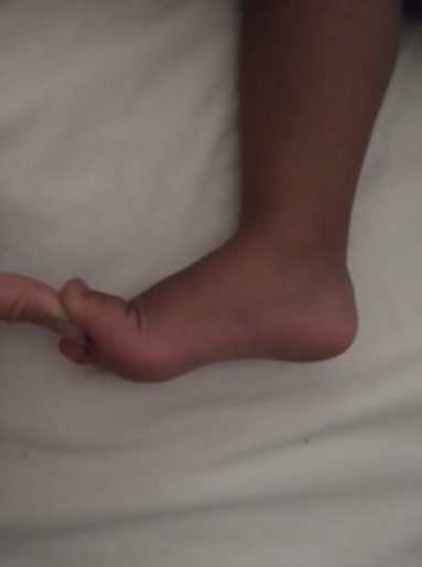
Clinical Paeds 12.png

Figure 12. Jacks test. The medial longitudinal arch is reconstituted with passive dorsiflexion of the hallux

A high arch is also examined with the child standing.  Observe the position of the heel from behind. The Coleman block test will distinguish a forefoot- from hindfoot- driven deformity. Again it is helpful to demonstrate the test.

  • Ask the child to stand barefoot on a block. 
  • Adjust the position of the foot so that the hind foot and lateral border remain supported but the first ray is allowed to drop. 
  • Observe the position of the heel from behind.  The block test will accommodate a forefoot driven deformity and heel valgus should be restored. Residual varus suggests fixed deformity of the subtalar joint.

Looking at the posture of a parents foot can give significant information in the case of possible hereditary diagnosis such as Charcot-Marie-Tooth disease

Remember to watch the child walk and look for aids or orthotics.

When sitting palpate the foot for areas of tenderness.  Common paediatric sites of pain include those from the osteochondroses; over the heel, the navicular or second metatarsal. 

Forefoot deformities should be examined for degree of correctability and range of movement at each joint should be noted. 

Range of movement at the ankle and hind foot is recorded.  Silfverskiöld test will demonstrate the role of gastrocnemius in any equinus contracture.

  • With the knee fully flexed maximally dorsiflex the ankle.
  • Ensure the foot doesn’t fall into valgus as this will give a false impression of maximum dorsiflexion.
  • Slowly extend the knee, isolated gracilis tightness will result in reduced dorsiflexion of the foot as the knee extends.
 Clinical Paeds 13(a).pngClinical Paeds 13(b).png
 
Figure 13. Silfverskiöld test 

Cavus deformities, clawing of the toes or asymmetry may suggest neurolomuscular aetiology. Full examination of the central or peripheral nervous system should be performed where appropriate 

Examine individual muscle strength and grade according to the MRC grading system. 

Remember to examine the spine for stigmata of dysraphism

Table 3.png

Table 3.MRC Grading System ref: Medical Research Council. Aids to the examination of the peripheral nervous system, Memorandum no. 45, Her Majesty's Stationery Office, London, 1981

Examination of the shoulder

With the child standing and appropriately undressed examine the shoulder contour from the front, the side and from the back.  From the front facial features associated with dysplasia may also be observed.  Drooping of the mouth may be seen with fascioscapular humeral dystrophy.  Chest wall deformities may be seen with Poland’s syndrome or Marfan’s syndrome. 

From the back, winging or failure of scapular descent may be seen.   Shortening of the neck with a low hair line indicate abnormalities of vertebral segmentation as in Klippel-Feil syndrome.

Palpation of the bony contours identifies any areas of tenderness, swellings or deformity.  Pseudoarthroses of the clavicle may be identified or callus from historical fracture.

Range of motion should be examined and importantly functional limitations.  Can the child bring their hand to their mouth, to the back of their head, and behind their back?

Clinical Paeds 14(a).pngClinical Paeds 14(b).pngClinical Paeds 14(c).png

Figure 14.  Screening of shoulder range of motion

Teenagers may present with multidirectional instability with associated ligamentous laxity.  Habitual dislocators may be able to demonstrate signs spontaneously. 

Look for structural instability that may warrant further investigation.  High demand training in younger adolescence means that injuries previously seen in adults are seen at a younger age. 

Examination of the Elbow 

Post-traumatic deformities are commonly seen in the paediatric elbow including the gunstock deformity of a malunited supracondylar fracture. Examine the carrying angle with the child standing and the arm fully extended. Cubitus valgus following a lateral condyle fracture may present with ulnar nerve symptoms and indicate the need for a neurological examination. 

The true carrying angle will not be seen if there is fixed flexion deformity.  Range of flexion and extension is possibly best assessed with the arms fully abducted and quickly screens upper limb function.

Rotation is examined with the elbow flexed at the side of the body. Demonstration of rotation holding a pen or with the thumb extended is easiest for a child to follow. Restriction of rotation may reflect synostosis or congenital radial head dislocation.

Clinical Paeds 15(a).pngClinical Paeds 15(b).png

Figure 15. Restriction of left forearm pronation

Palpation of the bony anatomy may reveal incongruency of the radio-capitellar joint.  Comparison to the contralateral arm is essential and may help differentiate from a missed monteggia lesion. 

Look at the hands and knees for evidence of nail patella syndrome and for signs of ligamentous laxity. 

Examination of the hand and wrist

Observation plays a significant role in the examination of the hand in the child.  Congenital anomalies of the fingers may be isolated or part of a generalized syndrome.  Engage with the child using toys that require dexterity, looking at functional use of the hand or neglecting to use the limb.  

Inspecting more closely may reveal deformity, muscle wasting, swellings or contractures. Examine the range of movement and flexibility. 

Common deformities include clinodactyly, camptodactyly, polydactyly and syndactyly. Syndactyly may be simple or complex.

Preaxial longitudinal deficiencies including radial club hand should prompt investigation of the cardiac and renal systems.

Swellings of the hand and wrist, as with elsewhere, should be differentiated by their tissue of origin. Examples include epidermal inclusion cysts, ganglia, giant cell tumour, enchondroma or exostosis.  Synovitis may suggest an underlying inflammatory arthropathy.  

Prominence of the distal ulna with radial and volar deviation of the wrist is the classic Madelung deformity with physeal arrest of the growth of the distal radius.  It may be familial and part of a spectrum of dysplasia including Leri-Weill syndrome.  Is the height normal? Or it may be the consequence of traumatic injury.

Examine range of movement of the wrist and palpate or areas of tenderness.

Neurological examination in a child can be difficult but it may be important to document the function of the peripheral nerves.  Asking a child to make ‘a star’, ‘the ok sign’ and a ‘thumbs up’ screens the motor function of the ulnar, anterior interosseous and radial nerves respectively.  At the least, rolling a pen over the skin can indicate autonomic function of the nerves in very young children

Clinical Paeds 16(a).pngClinical Paeds 16(b).pngClinical Paeds 16(c).png

Figure 16. Neurological examination of the nerves of the upper limb

Neuromuscular examination

The approach to examining a child with cerebral palsy or other neuromuscular condition depends very much on their level of function.  If the child is ambulant observe them standing and walking. Alternatively examination begins in the wheelchair followed by supine on the couch.   

With time muscular imbalance leads to contracture and fixed deformity.  Examination of the lower limbs in the neuromuscular patient follows a sequence of joint examination looking for degree of contracture and spacticity. 

On standing, hip joint contracture can result in increased pelvic tilt and lumbar lordosis. There may also be pelvic tilt in the coronal plane associated with a scoliosis. 

Gait abnormalities range from tiptoe walking to crouch and scissoring as the child becomes heavier and contractures increase.   

In children with cerebral palsy prone examination is very informative.  Staheli’s test, described above, gives an accurate assessment of hip flexion contracture. 

Duncan Ely test looks specifically at rectus femoris tightness.  With the child prone flex the knee, if the buttock rises from the couch the rectus femoris is tight.

Supine, square the pelvis and perform Thomas’s test. 

In the younger child adduction contractures are best examined with both hips flexed and abducted simultaneously. Symmetry and combined abduction arc can be easily visualized. 

Phelp’s test is performed to examine contracture of gracilis.

  • With the child supine, flex the knee with the hip fully abducted.
  • The knee is then gradually extended.
  • Tightness of gracilis will result in adduction of the hip as the knee extends
Clinical Paeds 17.png
 
Figure 17. Phelps test. With tightness of the gracilis, the hip will adduct as the knee is extended.

Examination of the popliteal angle measures the degree of hamstring tightness.  The angle (residual popliteal angle) is made between the front of the leg and the vertical when the hip is fully flexed and the knee in maximum extension.

Clinical Paeds 18.png

Figure 18. Examination of the popliteal angle.

Silverskiolds test demonstrates isolated gastrocnemius tightness.

  • With the knee fully flexed maximally dorsiflex the ankle.
  • Ensure the foot doesn’t fall into valgus as this will give a false impression of maximum dorsiflexion.
  • Slowly extend the knee, isolated gracilis tightness will result in reduced dorsiflexion of the foot as the knee extends

Torsional deformities result from muscular imbalance and rotational profile examination is important to document.

Examination of the spine in a child 

Examination of the spine begins with standing.   Look for stigmata of dysraphism, or skin lesions associated with neurofibromatosis.  From the side observe for normal sagittal contours, excluding excessive kyphosis or lordosis.   From the back assess the height of the shoulders and any asymmetry of the flank creases. A plumb line dropped from the vertebrae prominens should pass through the centre of the sacrum.

Clinical Paeds 19.png

Figure 19.  Adolescent idiopathic scoliosis

A structural scoliosis can be differentiated from a postural scoliosis with Adams forward bend test.

The patient is asked to bend forwards revealing an asymmetrical rib or lumbar hump. 

Lifting them from under their arms, allowing body weight to elongate the spine, can assess flexibility of the curve in a very young child.  In an older child side bending is helpful but standing radiographs are required for true assessment of flexibility.

Clinical Paeds 20.png

Figure 20.  A rib hump seen on Adams forward bend test.

Feel for tenderness, assess straight leg raise and popliteal angles for hamstring tightness associated with spondylolysis or spondylolisthesis.  

Perform a full neurological examination where indicated

Previous
Next