TOPIC DETAILS

  Team Member Role(s) Profile
Paul Banaszkiewicz Paul Banaszkiewicz Section Editor
Rebecca Rebecca Mazel Segment Author
Nicole Nicole Abdul Segment Author
  • Benign

Fibrous cortical defect (non-ossifying fibroma) 

Incidence 

  • A developmental defect in which a nest of fibrous tissue appears in the bone and persists for several years.
  • 20% of benign bone lesions.
  • Male more than female.

Clinically 

  • Usually an incidental finding in children.
  • Most heal spontaneously.
  • Larger ones may ®pathological fracture (common presentation).

X-rays 

  • Lucent 
  • in the cortex of a long bone.
  • Metaphyseal 
  • Eccentric 
  • Margin well defined,sometimes scalloped and often sclerosed.
FT1.pngFT2.png
Case courtesy of Dr Jeremy Jones, <a href="https://radiopaedia.org/">Radiopaedia.org</a>. From the case <a href="https://radiopaedia.org/cases/23651">rID: 23651</a>
 
Histology
  • Whorled fibrous tissue, foam cells.
  • Occasionally, small elongated giant cells.

Differential diagnosis

  1. Malignant fibrous histiocytoma 
  2. Eosinophilic granuloma 
  3. Osteosarcoma
  4. Histiocytic lymphoma 
  5. Pyogenic osteomyelitis 

CASE BASED DISCUSSIONS

Case based Discussion Non Ossifying Fibroma 

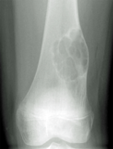
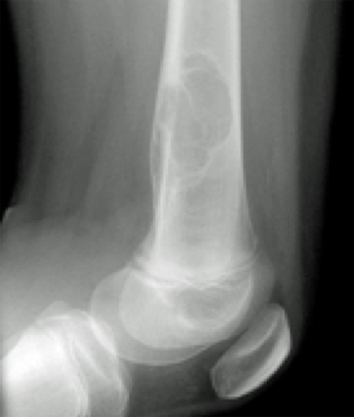
An 11y old girl twists her knee playing football. She attends A&E and has the following X-rays

Examiner:Please describe the X-rays 
NOF1.png

 NOF2 .png

Candidate:Distal femur metaphysis. Multiloculated lucent lesion with a sclerotic rim. Well defined/narrow zone of transition. No visible cortical erosion or soft tissue component. Extending to >50% of the width of the bone. These X-rays are insufficient to assess her twisted knee injury. 

Examiner:What are the differentials?

Candidate:

  • Non ossifying fibroma 
  • Malignant fibrous histiocytoma 
  • Eosinophilic granuloma 
  • Osteosarcoma 
  • Histiocytic lymphoma 
  • Pyogenic osteomyelitis 
Previous
Next

QUESTION 1 OF 3

Concerning non ossifying fibroma

QUESTION ID: 31

1. A peak incidence at 10-15 years of age
2. Are best treated with prophylactic nailing if they take up more than 50% of the width of the bone
3. Is an benign aggressive tumour
4. Is more common on the diaphysis
5. Will always require a biopsy to definitively diagnose

QUESTION 2 OF 3

The histological appearances of a non ossifying fibroma include

QUESTION ID: 32

1. Densely packed giant cells
2. Multipotent stromal cells
3. Prominent collagen formation
4. Storiform pattern of fibroblasts
5. Uniform small round blue cells

QUESTION 3 OF 3

A 10-year-old boy presented with a painful right hip. X-rays reveal a lesion in the proximal femur with loss of the normal trabecular architecture and a ground glass matrix within it.
Which of the following statements is not true:

QUESTION ID: 1095

1. A. Fibrous dysplasia is a mosaicism, meaning it can be polyostotic in one limb or one side of the body
2. B. Large lesions, particularly in the proximal femur may cause fractures
3. C. Patients may have an elevated blood level of FGF 23
4. D. Polyostotic fibrous dysplasia can be associated with hormonal abnormalities in McCune Albright syndrome
5. E. Polyostotic fibrous dysplasia has an autosomal dominant inheritance