TOPIC DETAILS

  Team Member Role(s) Profile
Paul Banaszkiewicz Paul Banaszkiewicz Section Editor, Segment Author
Nicole Nicole Abdul Segment Author
  • Recognise matrix as woven bone (osteoid).
  • Stroma: 

Fibrovascular = benign.
Sarcomatas = malignant.

  • Benign
  • Epiphyseal

None characteristic

  • Metaphyseal and diaphyseal

Osteoid osteoma and osteoblastoma have similar histology, but different clinical, radiological, and gross pathological findings. 

  • A small (<2 cm), benign, solitary, painful lesion of bone seen mainly in children and adolescents. 

Aetiology 

  • Arise from osteoblasts but the cause is unknown.

Incidence 

  • Accounts for 10% of benign bone tumours.
  • Third most common benign bone tumour.
  • Male:Female – 3:1.
  • Peak age 5–25 years (85% in this range).
  • Rare over 40 years of age.

        Location: any bone, rarely multifocal:

  • Tibia and proximal femur in 50%.
  • Spine (posterior elements).
  • Only occurs in bones formed by endochondral ossification.
  • May affect any part of a bone but it is usually intracortical.

Clinical signs 

  • Dull painis the most common presentation.
  • Pain often worse at night and relieved by NSAIDs.
  • Produce high levels of PGE2(this may account for why aspirin inhibits pain).
  • 10% occur in the spine and may lead to scoliosis.
  • In other sites may lead to: joint effusion, localised increase in temperature, synovitis, bone deformity, muscle atrophy.
  • Epiphyseal lesions can cause growth disturbance.
  • Runs a self-limiting course but usually surgery is required for pain relief.
  • Pain usually decreases as the lesion matures lasting 18–30 months.
  • Lesion healed by 3–7 years.

Imaging

  • The classic presentation on radiographs is a 2 cm (or smaller) round/oval lesion with a homogeneous dense centre and a peripheral radiolucent zone.
  • A lytic nidus may be seen surrounded by dramatic reactive sclerotic bone (which may mask the nidus).
  • CT scan is the gold standard imaging for diagnosis.
  • CT appearance is of a small, well delineated, low attenuation nidus surrounded by a dense sclerotic reaction.
  • MRI shows marrow and soft tissue oedema which can mask the nidus unless gadolinium is used to enhance it.
  • The tumour has intermediate intensity on T1 images. The nidus and oedema have a high intensity on T2 images (which is why the nidus can be obscured).
  • Hot spot on bone scan.
  • Bone scan usually demonstrates an intense area of radiotracer uptake in the region of the nidus and less in the reactive bone. This pattern, which is known as the double-density sign, is diagnostic of osteoid osteoma.

Differential diagnosis 

  1. Bone island (enostosis).
  2. When small in size a Brodie’s abscess may appear similar to an ostoid osteoma on plain radiographs.
  3. Osteoblastoma – although these are larger in size and while painful do not have the characteristic night exacerbation. They also have a predilection for vertebrae.
  4. Stress fracture.
  5. Eosinophilic granuloma.
  6. Intracortical haemangioma.
  7. Ewing’s sarcoma (particularly on MRI images).
  8. Intacortical osteosarcoma.
  9. Bone abscess.
  10. Osteomyelitis.

Pathology

  • Nidus is usually a well defined red coloured area, less than 1 cm in diameter.
  • Osteoid osteomas histologically have:
    • An inner nidus of blood vessels, connective tissue, osteoblasts, osteoid and woven bone. Osteoclasts may also be present. The nidus may be calcified.
    • Reactive sclerosis is the outermost part of the tumour


BSBONETUMOUROSTEOGENIC 1(a).jpg


BSBONETUMOUROSTEOGENIC 1(b).jpg

Figure 1(a) and (b).There is diffuse thickening of the anterolateral distal femoral diaphyseal cortex with loss of corticomedullary differentiation as well as expansion. No aggressive periosteal reaction is identified. No discrete lucency or associated soft tissue mass.

 BSBONETUMOUROSTEOGENIC 2.jpg

Figure 2. There is dense right distal femoral diaphyseal cortical thickening with a central nidus. Patchy hyperostosis within the medullary cavity. No adjacent soft tissue abnormality.

BSBONETUMOUROSTEOGENIC 3.jpg

Figure 3. Osteoid osteoma  located largely within the thickened cortex and partially in the medullary cavity.

BSBONETUMOUROSTEOGENIC 4.jpg

Figure 4. Histology .A small central nidus composed of bony trabeculae (right two-thirds of this image) surrounded by a zone of sclerotic bone (left one-third of the image). The nidus and surrounding bone are sharply demarcated from each other. The osteoid trabeculae are separated by vascular fibrous connective tissue.

Treatment

NSAIDs:

  • Relieve symptoms.
  • May take 3–4 years for symptoms to resolve.

Surgical: 

  • Nidus excision – definitive treatment without recurrence (needs only intact rim of reactive bone around the nidus to ensure complete excision, this is called en blocexcision).
  • Localisation of the lesion with: 
  1. Bone scan (preoperative).
  2. Tetracycline (4 mg tetracycline per kg QDS PO 1–2 days preoperatively – the osteoma is then excised under UV light).
  3. CT (preoperative).
  4. X-ray excised tissue containing the nidus (intraoperative).
  • Beware of the dumbbell shaped nidus – make sure all of the osteoid osteoma is excised.

Percutaneous radiofrequency ablation/coagulation:

  • While the patient is under general anaesthetic, a CT scan is used to localise the nidus.
  • A needle is inserted into the nidus percutaneously under CT guidance.
  • A biopsy confirms correct localisation of the lesion.
  • A radiofrequency electrode is introduced into the nidus via a cannula. The electrode reaches a temperature of 90°C for 6 minutes.
  • The procedure is more than 90% successful.


  • Sometimes called a giant osteoid osteoma as they are histologically very similar.

Incidence: 

  • Approximately 1% of primary bone tumours.
  • Peak age: second/third decade of life. 80% of patients are less than 30 years old.
  • Predilection to male individuals.

Clinical signs 

  • Less intense pain than osteoid osteoma which is often not relieved by aspirin.
  • The vertebrae and long bones such as the femur and tibia are frequently affected.
  • May occur in pelvis or phalanges.
  • When the lesion is in the spine, it is often associated with scoliosis and there may be neurological signs.
  • Decreased range of motion in area affected.
  • Localised swelling.

Imaging

On X-ray:

  • Normally >2 cm in size.
  • Well defined lyticlesion, sometimes with areas of calcification.
  • Thin edge of reactive sclerosis around lytic lesion but less than in an osteoid osteoma.
  • Expansive – associated with cortical expansion and sometimes resultant destruction of the cortex.
  • There may be associated: periostitis, sclerosis, a soft tissue mass, or an aneurysmal bone cyst.
  • MRI tends not to be a useful investigation at looking at osteoblastomas in detail as often the soft tissues around the lesion enhance as well, even with gadolinium.

Differential diagnosis 

  • Osteoid osteoma.
  • Giant cell tumour.
  • Osteosarcoma (if more aggressive).

Pathology 

  • As for osteoid osteoma but increased size (2–10 cm).
  • Texture is gritty and friable.
  • Histologically the nidus consists of bone trabeculae surrounded by highly vascularised fibrous tissue.
  • Active osteoblasts line the periphery of trabeculae.
  • The nidus is surrounded by a thin zone of reactive bone/periostium.
  • Malignant change has been reported.
  • There is a spectrum of aggressiveness but they never metastasise.

Treatment 

  • Intralesional resection and cementing/bone graft to fill the defect in the bone.
  • Cryotherapy (PMMA) is used as an adjuvant.
  • En blocresectionfor large lesions or lesions in the spine. These can sometimes recur.
  • Radiotherapy may be given after resection.

Prognosis 

  • Recurrence ususally occurs within 2 years of treatment but most patients do not have any further lesions after the primary treatment.
  • Recurrence is more common in the spine.
  • Tumours can be aggressive locally but do not metastasise.

Comparison


Osteoid osteoma

Osteoblastoma

Incidence 

Common 

Rare 

Clinical 

Pain relieved by aspirin 

Pain partially/not relieved by aspirin 

Pathology 

Nidus <1–2 cm; woven bone; surrounded by reactive bone 

Nidus 3–10 cm; sheets of woven bone surrounded by thin zone of reactive bone

X-Ray 

Small central nidus surrounded by dense reactive bone 

Lucent or dense lesion 

  • Malignant
  • Epiphyseal
    • None characteristic
  • Metaphyseal and diaphyseal


  • Primary malignant tumour arising from mesenchymal cells and producing malignant osteoid. 

Incidence:

  • Affects about 1/200,000 population.
  • Accounts for 21% of malignant primary bone tumours (most common primary bone sarcoma).
  • More common in male individuals.
  • Bimodal age distribution:

Peak 1: 10–20 years (age of rapid growth).

Peak 2: 50–70 years (80% less than 30 years and those more than 40 years usually secondary to Paget’s).

  • Third most common malignancy in adolescents, after leukaemia and lymphoma.
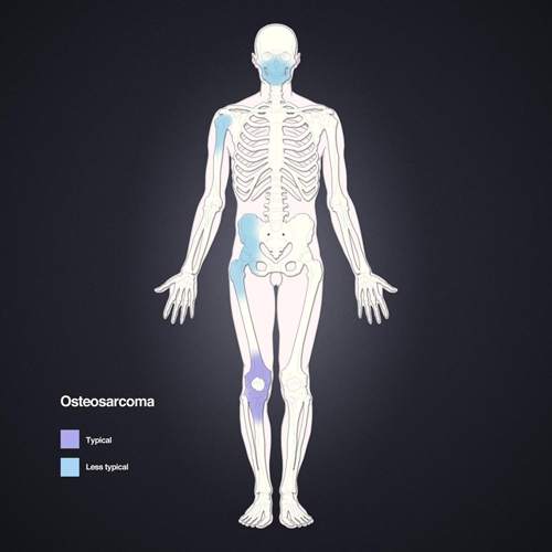
  • Most common location is in distal femur/proximal tibia but other sites include: humerus, skull, jaw, and pelvis.

Types 

Intramedullary (classic or ordinary) osteosarcoma.

Surface osteosarcomas: 

  • Parosteal osteosarcoma
  • Periosteal osteosarcoma
  • Intracortical osteosarcoma

Secondary osteosarcomas: 

  • Paget’s
  • Post radiation

Telangiectatic osteosarcoma.

BSBONETUMOUROSTEOGENIC 5.jpg

Figure 5.  Osteosarcoma is mainly seen in bones with the fastest rates of growth such as long tubular bones of the limbs.

Clinical signs

  • Pain which is worse at night.
  • May have a painful, localised swelling without a definite edge, which can be attached to surrounding soft tissues.
  • If vascular the mass may pulsate and feel warm.
  • A fracture following minimal trauma, if secondary to osteosarcoma, is often a late feature of these tumours.
  • Alkaline phosphatase is often greatly increased on blood test results.

Imaging 

  • X-rays:
  • Commonly show bone destruction (blastic, destructive lesions) with associated cortical breach.
  • Sun ray spicules/hairs on end patterns on radiographsand Codmans triangle(both caused by periosteal reaction) may be present.
  • Moth eaten/fluffy appearance of bone on radiographs.
  • A soft tissue mass may be visible.
  • Joint spaces are rarely involved.
  • The matrix produced by the tumour may be ossified/calcified in places.
  • MRI is used to evaluate the tumour in detail, including staging.
  • CT does not give any more information to MRI/plain radiographs apart from in lytic lesions where it can identify areas of mineralisation.

Pathology 

  • Pleomorphic and anaplastic cell population with abundant fibrous and chondroid matrix..
  • Stroma of spindle cells with numerous mitoses.
  • Most are high grade aggressive tumours approximately 10 cm in diameter at diagnosis.
  • 50% – osteoblastic 
  • 25% – chondroblastic
  • 25% – fibroblastic 
  • Associated with areas of high osteoblastic activity e.g. the metaphysis, childrens’ bones, and bones affected by Paget’s disease.
  • 20% are secondary to other conditions (e.g. Paget’s disease, enchondromas, osteochondromas, chronic osteomyelitis, irradiation, fibrous dysplasia, osteopetrosis and bone infarction).
  • Usually occurs in the metaphysis and initially extends within the medulla but quickly perforates the cortex and causes a periosteal reaction.
  • As the tumour mass expands new bone forms along vascular channels giving the appearance of sunray spicules.
  • Metastasises via the blood stream to the lung and other bones.
  • Carriers of the retinoblastoma tumour suppressor gene are at a high risk of developing osteosarcomas.
  • In patients with Rothmund Thomson syndrome, there is a defect in the RECQL4gene on chromosome 8. 
  • These patients also are at high risk of developing osteosarcomas.
BSBONETUMOUROSTEOGENIC 6(a).jpg

BSBONETUMOUROSTEOGENIC 6(b).jpg


Figure 6 (a) and (b). AP and lateral radiographs left tibia. Classic osteosarcoma.A sclerotic lesion involving dia-metaphyseal region of the tibia with a wide zone of transition, osteoid matrix, periosteal elevation (Codman’s Triangle) and characteristic " Sunburst " type of periosteal reaction.

BSBONETUMOUROSTEOGENIC 7(a).jpg

BSBONETUMOUROSTEOGENIC 7(b).jpg

Figure 7. Fracture distal (metadiaphysis) femur with overriding and posteromedial displacement of the distal fracture segment. Small laterally displaced osseous fragment is also seen. The fracture margins are irregular. There are ill-defined lucencies reflective of lytic changes involving the distal shaft, metaphysis and epiphysis of the femur. Interrupted periosteal reaction (Codman triangle)is seen at the distal femoral shaft above the fracture site. Lobulated soft tissue mass density is seen surrounding the fracture and effacing the adjacent fat planes. Most important for candidates is too realise that this is a pathological fracture and not a de nova fracture.

Differential diagnosis 

  • Post traumatic callus or myositis ossificans.
  • Stress fracture - pathology may look similar.
  • Osteomyelitis or syphilis
  • Benign bone tumour may have a similar appearance especially if X-rayed early.
  • Ewing’s sarcoma
  • Bone metastasis
  • Aneurysmal bone cyst

Treatment 

  • T10 regimen (methotrexate, vincristine, adriamycin) has approximately 60–75% survival.
  • 45% of patients with the T10 regimen achieve a 100% kill rate and therefore 100% survival.
  • Survival beyond 10 years is regarded as cured.
  • Regimen continued postoperatively or changed to cisplatin depending on histology ®92% of patients are disease free at 2 years.
  • Chemotherapy continues for 12 months in 4/52 cycles.
  • Intra-arterial chemotherapy is used in some centres. This is when increased doses of the drugs are given at the site of the lesion but there is no evidence that this changes the outcome as multiple feeding vessels of the tumour may be missed.
  • Radiotherapy is used for palliation of local pain and to treat surgically inaccessible lesions and painful metastatic deposits.
  • Osteosarcomas are relatively radio-resistant but radiotherapy may also be used preoperatively to decrease the size and vascularity of the tumour.
  • Prophylactic irradiation of the chest has not been shown to be effective.

Surgery:

  • Aggressive resection including limb amputation with adjuvant chemotherapy may be necessary.
  • Neoadjuvant chemotherapy may be used for a number of weeks before surgery is attempted as a limb salvage procedure. Postoperative chemotherapy is also required in these cases.
  • Limb salvage requires ability to: 
  1. Preserve nerves.
  2. Preserve or reconstruct vessels.
  3. Preserve sufficient muscle for functional motor power and soft tissue coverage.
  4. Achieve safe, tumour-free margins (wide).
  • Reconstruction options: 
  1. Allografts
  2. Endoprosthesis
  3. Expendable bone (fibula, ilium)
  4. Rotationplasty

Prognosis 

  • Untreated cases have a 95% death rate in 2 years.
  • 10% have macro-metastases at presentation; 90% have micro-metastases.
  • Even with metastatic (stage 3) disease,5 year survival now is 30–40% (10–20% with surgery alone).

Prognostic factors: 

  • Age – adults do worse.
  • Size of primary tumour (big is bad).
  • Location (proximal worse than distal).
  • Type:

Parosteal – tend to be more low-grade tumours and therefore these have a better prognosis.

Intraosseous (classic) osteosarcomas have a good prognosis.

  • Stage
  • Response to chemotherapy:

80–90% 5-year disease-free survival in good response patients.

60% 5-year disease-free survival in patients with poor response rates.

  • More than 16 metastatic deposits is considered to have a poor prognosis.
  • Pathological fracture(s) do not affect prognosis.
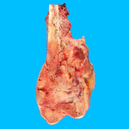
BSBONETUMOUROSTEOGENIC 8.jpg

Figure 8.Gross pathology distal femur.
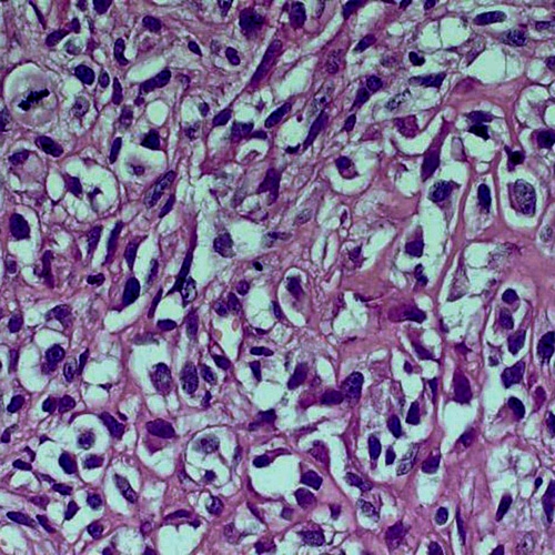

BSBONETUMOUROSTEOGENIC 9.jpg

Figure 9. In addition to mitosis (upper left) and cellular atypia (everywhere), which are usual; one should see the atypical osteoid (upper right) to make the diagnosis.
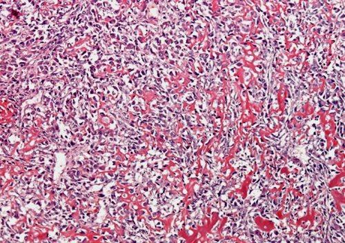
BSBONETUMOUROSTEOGENIC 10.jpg

Figure 10. Lace-like osteoid deposited in between heavily anaplastic tumour cells.

Incidence

  • 1% of primary malignant bone tumours.
  • Usually patients are older than 20 years old.
  • Peak incidence is 30–50 years.
  • Male:female 2:3

Clinical signs 

  • Usually presents with a constant ache or lump.
  • The lump may also be painless.
  • Usually occurs in long bones on the cortex of the metaphysis.
  • Usually presents as a stage 1A lesion (low grade).
  • Commonest site = posterior aspect of distal femur. Tumours at this site can cause a reduced range of motion.

X-rays 

  • Well circumscribed mass attached to cortex.
  • Often heavily ossified and may be lobulated.
  • May be separated from cortex by a lucent line (30%).
  • Broad-based tumour with mottled calcification.
  • The cortex is not eroded.
  • These tumours do not invade the medullary cavity (unlike chondrosarcoma).
  • CT scans are used for staging and to evaluate common sites of metastasis such as the lungs.
  • MRI is helpful in giving more information about the tumour and the extent of its spread into soft tissues.
  • Bone scan shows tumour site(s) as hot spots.

Pathology

  • Histologically the tumours look very similar to fibrous dysplasia.
  • Atypical spindle cells lie between normal osseous trabeculae.

Differential Diagnosis 

  • Osteochondroma
  • Myositis ossificans.
  • Defect in the bone at the adductor magnus insertion
  • Fibrous dysplasia (these can be excluded by looking at the radiographs as they present differently to parosteal osteosarcoma).

Treatment 

  • Chemotherapy or radiotherapy is not effective in preventing recurrence (although chemotherapy may be used as an adjuvant to surgery in high grade tumours). Wide surgical resection is the standard treatment.

Prognosis 

  • Said to be better than for classic osteosarcoma.
  • 70–80% 5-year survival.

Incidence

  • Rare 
  • Occurs in second to third decade of life.
  • Females>males

Clinical signs

  • Pain, swelling and tenderness at tumour site.
  • The midshaft of the femur and tibia are the most common location.
  • Patient’s first presentation may be following a pathological fracture.

Imaging

X-rays:

  • Ill-defined swelling
  • Sub-periosteal reaction giving a “hairs on end” appearance to the bone/swelling on bone.
  • Large external, poorly mineralised mass within a depression of cortical erosion.
  • The tumours do not normally extend into the medullary canal.

CT scan and bone scans are performed for staging.

Pathology

  • The tumour arises from the germinal layer of periosteum which causes a periosteal reaction above it.
  • Lobules of cartilage with malignant spindle cells is seen under microscope.
  • The tumour produces osteoid which is often found in between malignant spindle cells.
  • This type of tumour is characteristically a lower grade than classic osteosarcomas but a higher grade than parosteal osteosarcomas.
  • There may be areas of calcification and fibroblastic tissue.
  • Patients with these tumours may have a mutation of p53 gene.

Differential diagnosis

  • Periosteal osteosarcoma
  • Periosteal chondrosarcoma

Treatment

  • Neoadjuvant chemotherapy, surgical resection (limb salvage if possible), followed by postoperative chemotherapy.y

Prognosis

  • High affinity to metastasise to lungs (up to 35% rate).
  • 85–90% 5-year survival
  • Poor prognosis in tumours expressing multi-drug resistant gene portends due to chemotherapy not being effective.
  • Better prognosis than classic osteosarcomas, poorer prognosis than periosteal osteosarcomas.

Incidence

  • Age more than 45 years
  • Male:female 2:1 
  • Occurs in advanced polyostotic disease 10%.
  • Affects 1% of all patients with Paget’s.

Clinical signs

  • Uncontrollable pain
  • Common sites for these osteosarcomas include the pelvis, femur and humerus.
  • Many will already have metastasised to other sites by the time of diagnosis.

Imaging

  • X-rays of the bones affected and a bone scan should be done.
  • CT determines whether the disease has metastasised to the lungs.

Pathology 

  • 50% or more are osteosarcomas.
  • 25% fibrosarcomas
  • Remainder are chondrosarcomas or anaplastic tumours.

Treatment

  • Neoadjuvant chemotherapy, surgical resection, and post operative chemotherapy.
  • For tumours at sites where surgery is not possible (e.g. skull), chemotherapy and radiotherapy are used.

Prognosis 

  • Poor – less than 50% 1-year survival 

Criteria for diagnosis: 

  • Initial lesion must be benign in nature both histologically and radiologically.
  • Secondary malignancy must have arisen within the radiotherapy treatment zone.
  • A relatively long latent period must have elapsed before the clinical appearance of thesecondary lesion.
  • All secondary sarcomas must be identified histologically and different to that of the original pathology.
  • Mean latency to onset of 10 years.
  • Earlier appearance in younger patients.
  • In excess of 20 Gr radiation given.

Clinical signs

  • The spine, pelvis, hips and shoulders are common sites of these tumours.
  • Pain and swelling in a previously irradiated area of the body.

Imaging

  • Although a biopsy is needed for diagnosis, a CT scan can help stage the tumour and determine whether there have been any metastases (particularly pulmonary).

Treatment

  • As for other osteosarcomas: neoadjuvant chemotherapy, surgical resection, and post-surgical chemotherapy.

Prognosis 

  • Cumulative disease free survival rate = 17% at 5 years.
  • Location of the tumour can make surgical resection not possible and therefore prognosis is worse.

Incidence

  • Rare – 5% of all osteosarcomas.
  • More common in men.

Clinical signs

  • Aggressive form of osteosarcoma.
  • Pain, swelling and tenderness over tumour site.
  • Commonly present with a pathological fracture.
  • Arises within the diaphysis of long bones.
  • Femur and& tibia most common, then humerus.

Imaging 

  • X-rays:
    • Often entirely osteolytic 
    • Bone and cortex destruction (expansive)
    • Periosteal reaction
    • Codman's triangles (periosteum is lifted from bone due to periosteal reaction).
  • MRI should be performed to determine soft tissue involvement.
  • Bone scan

BSBONETUMOUROSTEOGENIC 11.jpg

Figure 11. AP radiograph distal femur demonstrating an aggressive lytic lesion in the distal metaphysis. Soft tissue swelling is noted. There are sparse areas of ossification in the tumour matrix.

 BSBONETUMOUROSTEOGENIC 12.jpg

Figure 12. MRI coronal T1 image.Multiloculated lytic lesion distal metaphysis femur. Multiple fluid-fluid levels are seen. Massive edema is seen in the muscular compartments. Bone marrow edema involves the epiphysis.

 BSBONETUMOUROSTEOGENIC 13.jpg

Figure 13. Telangiectatic osteosarcoma is a variant with distinct radiographic, gross, and microscopic features. On radiographs, it appears as a purely lytic expansile lesion with no evidence of sclerosis. Given the geographic bone destruction, it frequently presents with pathologic fractures. The gross specimens usually show an expansile mass composed of blood-filled cystic spaces separated by delicate septae. A fleshy solid component is not seen.

Pathology 

  • Gross appearance is a multi-cystic “bag of blood.”
  • Microscopically it has large blood filled spaces and thin septation. Within the septa there is scanty osteoid production by the pleomorphic malignant cells of this high grade tumour.

Treatment

  • As for other types of osteosarcoma: neoadjuvant chemotherapy, surgical resection, and postoperative chemotherapy.

Differential diagnosis

  • Aneurysmal bone cyst
  • Classic osteosarcoma

Prognosis 

  • Good if good response to chemotherapy.
  • Poor if multi-drug resistance gene portends are present on tumour.
  • Local recurrence is relatively uncommon but is associated with a poor prognosis.

Majority of the images courtesy of Radiopaedia found at https://radiopaedia.org

Previous
Next

QUESTION 1 OF 3

A 72-year-old man presents with increasing pain in the pelvis. A plain radiograph shows expanded bone in the superior pubic ramus, with coarse “purposeful” trabeculae. Further investigation with MRI shows an associated soft tissue mass arising from the area of abnormal bone.
Which of the following statements is true:

QUESTION ID: 54

1. A. Biopsy is not required
2. B. Bisphosphonate treatment has minimal role in the treatment of Paget disease
3. C. On isotope bone scan, the abnormal area in the pelvis is unlikely to show increased uptake
4. D. Osteosarcoma secondary to Paget disease has a different biological potential
5. E. The serum alkaline phosphatase is likely to be normal

QUESTION 2 OF 3

115.This 16-year-old  patient presents with pain on the upper medial side of his right leg when wearing his shin pads when playing football. Examination reveals a firm lump.(Figure 1)
Considering the presumed diagnosis which of the following statements is most accurate?

MHE.png lr.png

Figure 1 Clinical photo

QUESTION ID: 2207

1. Further examination could reveal radial head dislocation
2. He is likely to present later with genu varum.
3. The lump has a 2% chance of becoming malignant (5-10%)
4. This is most likely a subperiosteal haematoma
5. Transmission is autosomal recessive

QUESTION 3 OF 3

37.A 12-year-old boy presents with leg pain of 2 months. Radiographs show a periosteal reaction with histology reporting uniform small round blue cells.
Choose the genetic defect associated with this condition.

QUESTION ID: 2225

1. Mutation in epidermal growth factor protein (erbB-2) coding gene
2. Mutation of retinoblastoma protein (pRB-1) coding gene
3. Mutation of tumour protein 54 (p53) coding gene
4. Translocation t(11:22)
5. Translocation t(2:13)