TOPIC DETAILS

  Team Member Role(s) Profile
Paul Banaszkiewicz Paul Banaszkiewicz Section Editor
Rebecca Rebecca Mazel Segment Author
Nicole Nicole Abdul Segment Author

Features:

    • Haemangiomas are benign lesions characterised by vascular spaces lined with endothelial cells.
    • Common, with approximately 10% of autopsy cases having vertebral haemangiomas.
    • M:F 1:2
    • Age 30–50 years

Site

  • Vertebral bodies (thoracic especially) 50%.
  • Calvarium 20%
  • Remainder found in the tibia, femur and humerus.

Presentation

  • Usually asymptomatic and solitary discovered on X-ray or at post mortem.
  • Vertebral haemangiomas may present with chronic back ache and can cause neurological symptoms if they extend into the epidural space.
  • May present as a pathological fracture.
  • Long bones may over grow due to increased blood supply.

Radiology

Plain X-ray

  • Vertebral lesions have coarse, thickened vertebral trabeculae secondary to erosion of the horizontal trabeculae giving a “corduroy” appearance.
  • Vertical striations without bone expansion (differential diagnosis Paget’s(jail bar appearance).

 Vascular tumour 6.png


Computed tomography

  • Vertebral body lesions have a “polka dot” pattern as the vessels are seen in cross-section.
  • Calvarial lesions are lytic and resemble radiating wheel spokes.
  • Haemangiomas in the metaphysis or epiphysis of long bones are lytic lesions that give a spiculated pattern known as “Irish lace”.

Vascular tumour 3.pngVascular tumour 4.png


Vascular tumour 5.png

Magnetic resonance imaging

  • T1 sequences vary from low to high intensity depending on the amount of adipose tissue present.
  • T2 sequences demonstrate lesions with high signal due to the vascularity.

Pathology

  • Vascular hamartoma with cystic, dark red cavities.
  • Four types: capillary, cavernous, arteriovenous and venous.
  • Capillary and cavernous lesions are the most common in bone.
  • Arteriovenous haemangiomas are remnants of foetal capillary beds.
  • (Similar appearances = ABC, telangiectatic OS).

Microscopy

  • Non-vascular components of haemangiomas include fat, smooth muscle, fibrous tissue, bone, haemosiderin and thrombus.

Add Fig. 3

Treatment

  • Unnecessary unless the lesion is symptomatic.
  • Lesions in the calvarium should be resected with a thin margin of normal bone.
  • Vertebral lesions respond to radiation or can be treated with surgical excision preceded by embolisation.
  • Lesions in long bones should be excised and packed with bone graft if appropriate.
  • Seems to have two distinct clinical presentations.
  • First, the lesion can present as multiple lesions in a single bone, two or more adjacent bones, or perhaps all the bones of a limb.
  • These lesions seem to have an indolent course and the prognosis remains good.
  • The second presentation is that of single or multiple rapidly progressive lesions that metastasise to other bones or to the lung this form of the disease has a very poor prognosis.

CASE BASED DISCUSSIONS

Case based Discussion Haemangioma

A 45 year old woman had a spinal MRI for worsening chronic back pain. It shows an unobstructed spinal cord, but you notice a lesion on L1

Haemangioma spine 1.jpeg

Sagtital T2 image lumbar spine.High signal on T2 due to high fat content.

Examiner:What is a haemangioma?

Candidate:Haemangiomas are benign lesions characterised by vascular spaces lined with endothelial cells. They are common, usually asymptomatic, and found incidentally.

Examiner: Discuss the treatment options for this patient

Candidate:It is possible that the haemangioma is a contributing factor for her back pain. It could be treated with low dose radiotherapy, or embolisation then surgical excision. Long bone haemangiomas can be curetted and packed with bone graft.

Case based Discussion Renal Cell Mets

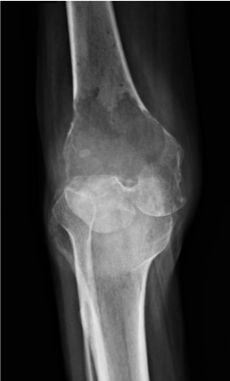
An active 62 year man presents to his GP with 8 weeks of pain in the left knee. He has tried physiotherapy but this has not helped. The GP refers him to a knee surgeon who performs the following X-ray
Vascular tumour 8.png

Anteroposterior(AP) radiograph knee

Examiner :The knee surgeon decides this warrants further investigation. Among other tests she orders a set of bloods. Which bloods would be appropriate?

Candidate:Standard FBC, U&E, coagulation screen. Bone profile to look especially for hypercalcaemia. Given the patient’s age, metastatic disease is high on the list of differentials. Tumour markers include CEA (bowel), Ca19-9 (pancreas), electrophoresis (myeloma), PSA (prostate), Ca125 (ovarian - if this was a female patient)

Examiner:What is Mirel’s score? How does it influence management?

Candidate:Mirel’s score is a set of criteria used to predict whether a bone containing a tumour will break. It assigns scores based on the lesion’s site, pain, size, and whether it is blastic or lytic. An increased score corresponds to an increased fracture risk.

Examiner:A CTCAP shows a mass in the left kidney, and renal cell carcinoma is diagnosed. Due to Mirel’s score, you feel that operative intervention is appropriate. Is there any procedure you would consider arranging prior to surgery? Which other diagnoses would you consider this for?

Candidate:Embolisation prior to surgery can reduce bleeding in highly vascular tumours such as renal, thyroid, and phaechromacytoma.

Previous
Next